Review
. 2020 Nov;127(11):1569-1577.
doi: 10.1007/s00702-020-02198-8. Epub 2020 May 8.

Do not keep it simple: recent advances in the generation of complex organoids

Affiliations
Review

Do not keep it simple: recent advances in the generation of complex organoids

Philipp Wörsdörfer et al. J Neural Transm (Vienna). 2020 Nov.

Abstract

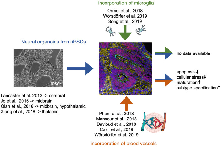
3D cell culture models which closely resemble real human tissues are of high interest for disease modelling, drug screening as well as a deeper understanding of human developmental biology. Such structures are termed organoids. Within the last years, several human organoid models were described. These are usually stem cell derived, arise by self-organization, mimic mechanisms of normal tissue development, show typical organ morphogenesis and recapitulate at least some organ specific functions. Many tissues have been reproduced in vitro such as gut, liver, lung, kidney and brain. The resulting entities can be either derived from an adult stem cell population, or generated from pluripotent stem cells using a specific differentiation protocol. However, many organoid models only recapitulate the organs parenchyma but are devoid of stromal components such as blood vessels, connective tissue and inflammatory cells. Recent studies show that the incorporation of endothelial and mesenchymal cells into organoids improved their maturation and might be required to create fully functional micro-tissues, which will allow deeper insights into human embryogenesis as well as disease development and progression. In this review article, we will summarize and discuss recent works trying to incorporate stromal components into organoids, with a special focus on neural organoid models.

Keywords: Blood vessel; Microglia; Neural; Organoid; Stroma; Vasculature.


Conflict of interest statement

The authors declare no competing interests.

Figures

Fig. 1
Fig. 1
Cellular composition of the nervous tissue. The nervous tissue consists of neurons and glial cells. Among the glial cells, astrocytes, oligodendrocytes, microglia, and ependymal cells can be found. Moreover, a stem and progenitor niche exists in close association with the ependymal cells as well as brain blood vessels. The surface of the brain tissue is covered by a layer of connective tissue including blood vessels, forming the meninges. While neurons, astrocytes, oligodendrocytes, and ependymal cells arise from a common Sox1+ neuroepithelial stem cell, the blood vessels and the meningeal connective tissue derive from the embryonic mesenchyme. In contrast, microglia are generated early during development in the yolk sac blood islands and subsequently infiltrate the nervous tissue to become brain tissue resident, self-maintaining macrophages. This schematic is composed of graphical elements taken from the image bank from Servier Medical Art licensed under a Creative Commons Attribution 3.0 Unported License (https://creativecommons.org/licenses/by/3.0)
Fig. 2
Fig. 2
Incorporation of microglia and blood vessels into neural organoids. This fig. shows recent publications describing (1) the generation of brain region specific neural organoids (Jo et al. ; Qian et al. ; Xiang et al. 2019), (2) the incorporation of microglia into neural organoids and (3) the incorporation of a vascular system into neural organoids as well as the so far demonstrated advantages of increasing organoid complexity. The phase contrast image (left side) shows undifferentiated iPSCs in 2D culture. The immunofluorescence image (right side) shows an immature neural organoid generated from hiPSCs stained for Sox1 (neural stem cells—> red), MAP2 (neurons—> yellow), and N-Cadherin (green). Graphical elements within this schematic were taken from the image bank from Servier Medical Art licensed under a Creative Commons Attribution 3.0 Unported License (https://creativecommons.org/licenses/by/3.0)

References

    1. Anderson KD, et al. Angiogenic sprouting into neural tissue requires Gpr124, an orphan G protein-coupled receptor. Proc Natl Acad Sci USA. 2011;108:2807–2812. doi: 10.1073/pnas.1019761108. - DOI - PMC - PubMed
    1. Asai A, et al. Paracrine signals regulate human liver organoid maturation from induced pluripotent stem cells. Development. 2017;144:1056–1064. doi: 10.1242/dev.142794. - DOI - PMC - PubMed
    1. Ayabe H, et al. Optimal hypoxia regulates human iPSC-derived liver bud differentiation through intercellular TGFB signaling. Stem Cell Rep. 2018;11:306–316. doi: 10.1016/j.stemcr.2018.06.015. - DOI - PMC - PubMed
    1. Bhaduri A, et al. Cell stress in cortical organoids impairs molecular subtype specification. Nature. 2020;578:142–148. doi: 10.1038/s41586-020-1962-0. - DOI - PMC - PubMed
    1. Bharadwaj S, et al. Multipotential differentiation of human urine-derived stem cells: potential for therapeutic applications in urology. Stem Cells. 2013;31:1840–1856. doi: 10.1002/stem.1424. - DOI - PubMed

Publication types

LinkOut - more resources