Review
. 2020 Sep;23(3):365-378.
doi: 10.1007/s40477-020-00468-5. Epub 2020 May 8.

Ultrasound of the ulnar-palmar region of the wrist: normal anatomy and anatomic variations

Affiliations
Review

Ultrasound of the ulnar-palmar region of the wrist: normal anatomy and anatomic variations

Stefano Bianchi et al. J Ultrasound. 2020 Sep.

Abstract

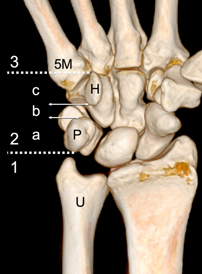
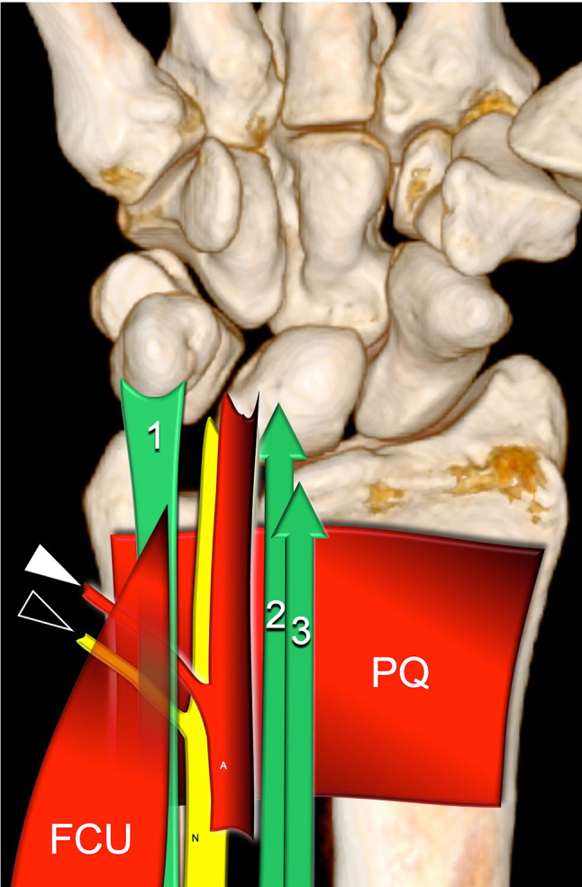
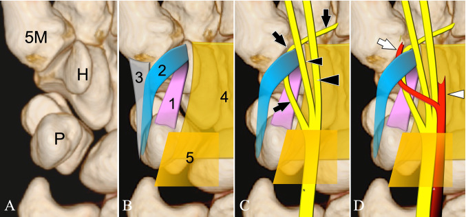
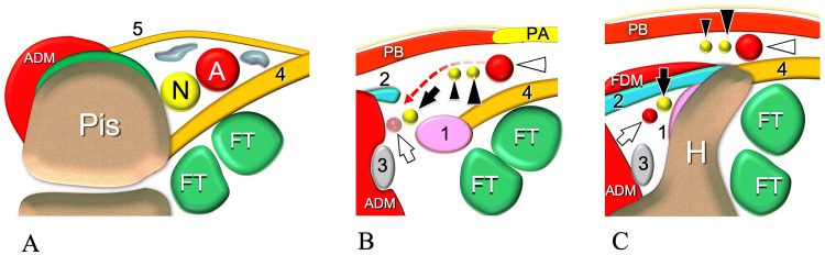
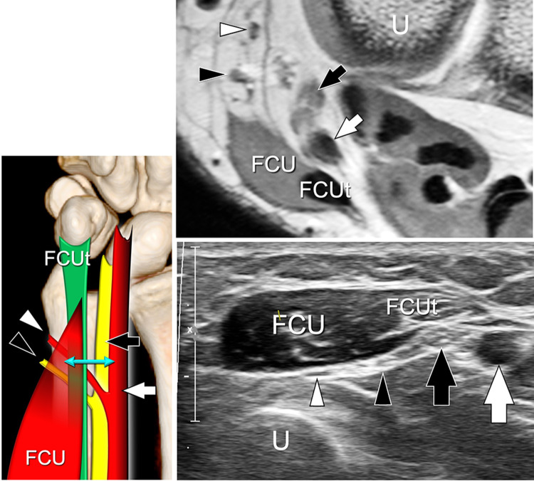
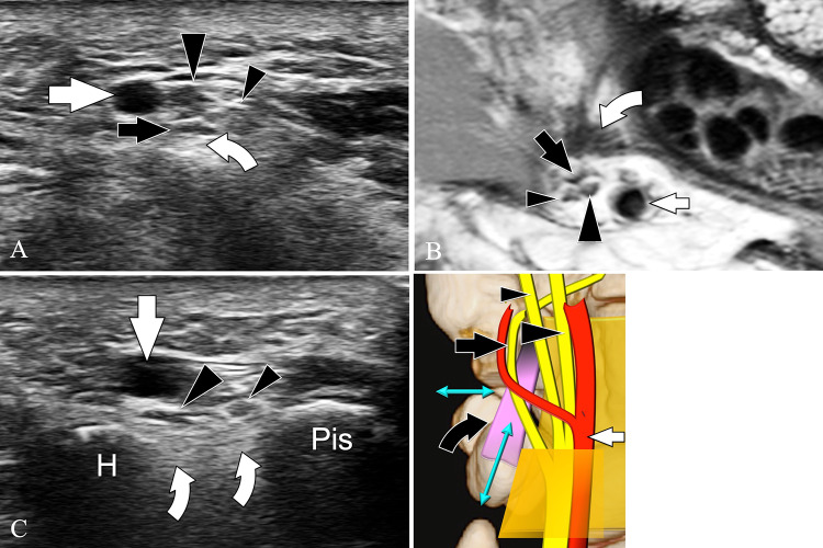
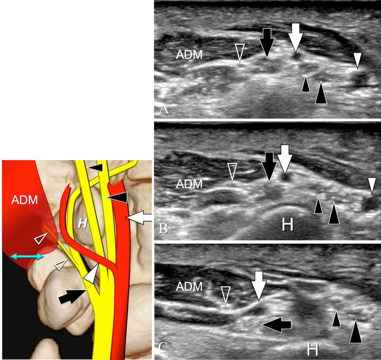
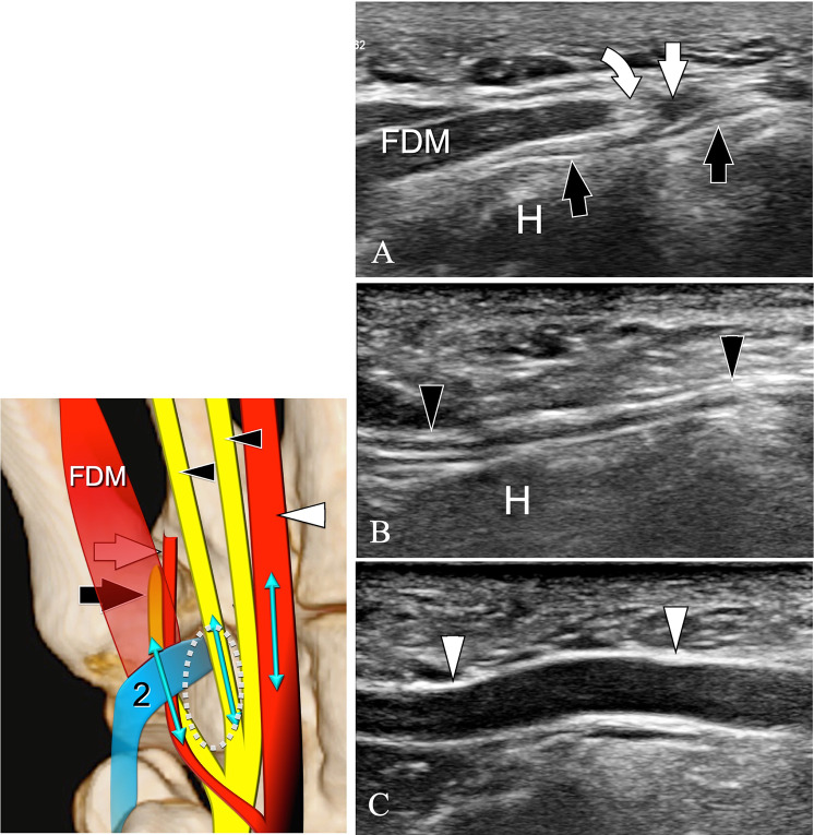
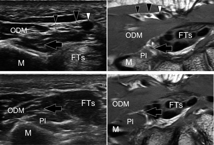
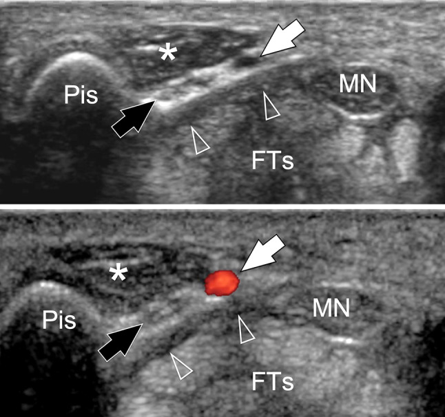
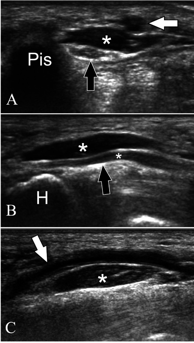
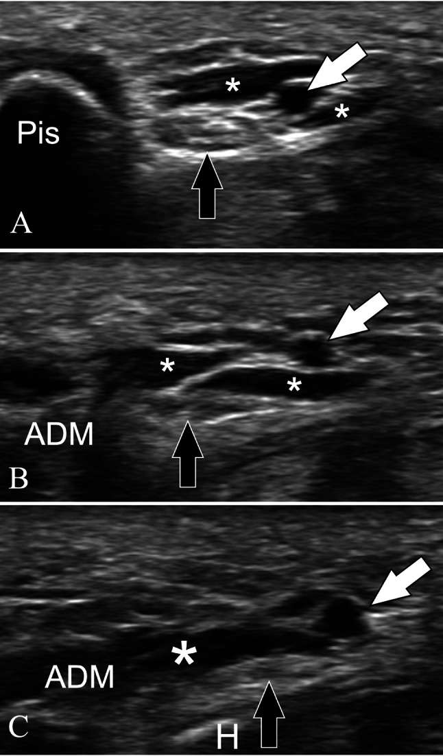
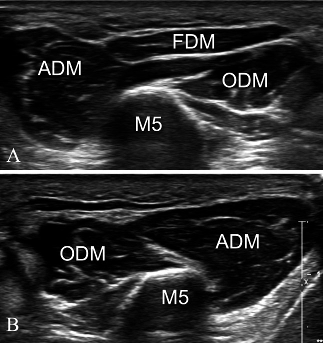
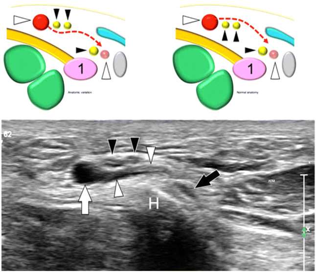
Ultrasound (US) assessment of the wrist is frequently used for the evaluation of carpal tunnel due to high frequency of local compression of the median nerve (MN), but the ulnar-palmar wrist region (UPWR) has received limited attention in the medical literature. The possibilities of US in the assessment of UPWR are therefore likely underestimated by sonologists. This review article is focused on the US assessment of the normal anatomy and anatomic variations of the UPWR. The anatomy of this region of the wrist is complex and less studied than the radial side. In an effort to simplify it and to present it didactically, we have divided this region in three parts on the basis of osseous landmarks. Our review indicates sonography is effective in identifying the UPWR and related disorders, and is thus a valuable tool for ensuring appropriate management of a variety of disorders.

Keywords: Guyon’s canal; Palmar carpal ligament; Transverse carpal ligament; Ulnar artery; Ulnar nerve.


Conflict of interest statement

The authors declare that they have no conflict of interest.

Figures

Fig. 1
Fig. 1
Palmar view of a reconstructed 3D CT of the wrist. Bone boundaries: fifth metacarpal (5 M), hook of the hamate (H), pisiform (P) and ulna (U). 1–3 correspond to the three regions as discussed in the text. For didactic purposes, the Guyon’s tunnel has been further divided in three parts (a–c)
Fig. 2
Fig. 2
The schematic drawing indicates the main anatomic structures of the first region (proximal to the pisiform). FCU = Flexor carpi ulnaris muscle and its tendon (1), PQ = pronator quadratus muscle. N = ulnar nerve and its dorsal cutaneous branch (black arrowhead), A = ulnar artery and its dorsal branch (white arrowhead), 2, 3 = flexor digitorum tendons of the fifth finger. Note that the dorsal branches of the ulnar nerve and artery run between the flexor carpi ulnaris and the pronator quadratus muscle
Fig. 3
Fig. 3
Schematic drawings (ad) indicate the main anatomic structures of the second region (Guyon’s tunnel). Coronal plane. a Bones: fifth metacarpal (5 M), hook of the hamate (H), pisiform (P). b Ligaments: pisohamate ligament (1), fibrous arcade of the hypothenar muscles (2), pisometacarpal ligament (3), transverse carpal ligament (4), palmar carpal ligament (5). c Nerves: Ulnar nerve (N) and its deep branch (black arrows). Proper digital nerve of the little finger (small black arrowhead) and the common digital nerve of the fourth webspace (large black arrowhead). d Arteries: Ulnar artery (A) and its deep (white arrow) and superficial (white arrowhead) branches
Fig. 4
Fig. 4
Schematic drawings (ac) indicate the main anatomic structures of the second region (Guyon’s tunnel). Sagittal plane. a Ligaments and bones: pisohamate ligament (1), fibrous arcade of the hypothenar muscles (2), pisometacarpal ligament (3). Fifth metacarpal (5 M), hook of the hamate (H), pisiform (P). b Nerves: Ulnar nerve (N) and its deep branch (black arrows). Proper digital nerve of the little finger (small black arrowhead) and the common digital nerve of the fourth webspace (large black arrowhead). c Arteries: Ulnar artery (A) and its deep (white arrow) and superficial (white arrowhead) branches
Fig. 5
Fig. 5
Schematic drawings indicate the main anatomic structures of the three parts of the Guyon’s tunnel. Axial plane. a, b drawings indicating the anatomy at the proximal (a), middle (b) and distal (c) parts. Bones: Pisiform (Pis), hook of the hamate (H). Muscles: abductor digiti minimi (ADM), palmaris brevis (PB) and flexor digiti minimi (FDM). Ligaments: pisohamate ligament (1), fibrous arcade of the hypothenar muscles (2), pisometacarpal ligament (3) transverse carpal ligament (4), palmar carpal ligament (5), palmar aponeurosis (PA). Nerves: Ulnar nerve (N). Deep branch of the ulnar nerve (black arrow). Proper digital nerve of the little finger (small black arrowhead) and the common digital nerve of the fourth webspace (large black arrowhead). Arteries: Ulnar artery (A). Deep (white arrow) and superficial (white arrowhead) branches of the ulnar artery. Tendons: flexor digitorum tendons of the fifth finger (FT)
Fig. 6
Fig. 6
Axial schematic drawings indicate the inlet and outlets (heavenly area) of the Guyon’s tunnel. The levels of the proximal hiatus (a) and of the two distal hiatus (b) are indicated in the coronal drawing by the two dotted lines (A and B). In b, the black asterisk indicates the pisohamate hiatus, while the white asterisk indicates the superficial hiatus. Bones: Pisiform (P), hook of the hamate (H). Muscles: abductor digiti minimi (ADM), palmaris brevis (PB). Ligaments: pisohamate ligament (1), fibrous arcade of the hypothenar muscles (2), pisometacarpal ligament (3) transverse carpal ligament (4), palmar carpal ligament (5). Nerves: Ulnar nerve (N) and its deep branch (black arrow). Proper digital nerve of the little finger (small black arrowhead) and the common digital nerve of the fourth webspace (large black arrowhead). Arteries: Ulnar artery (A) and its deep (white arrow) and superficial (white arrowhead) branches. Tendons: flexor digitorum tendons of the fifth finger (FT)
Fig. 7
Fig. 7
Schematic drawings indicate the main anatomic structures of the hypothenar region. Coronal plane. a superficial plane. Hypothenar fascia (HF), palmar aponeurosis (PA) palmaris brevis muscle (PB) b deep plane. Abductor digiti minimi (A) flexor digiti minimi (B) opponens digiti minimi (C), fourth palmar interosseous muscle (PIM). Nerves: Ulnar nerve (N) and its deep branch (black arrows). Proper digital nerve of the little finger (small black arrowhead) and the common digital nerve of the fourth webspace (large black arrowhead). Note the deep branch of the ulnar nerve entering the pisohamate hiatus (curved arrow), delimited by the pisohamate ligament (1), and the fibrous arcade of the hypothenar muscles (2)
Fig. 8
Fig. 8
Normal US anatomy of the UPWR. First region. Schematic drawing in the coronal plane. Axial sonogram and corresponding T1-weighted MR image obtained at the level of the double arrow. Ulnar nerve (black arrows) and its dorsal cutaneous branch (black arrowheads), ulnar artery (white arrows), and its dorsal cutaneous branch (white arrowheads). 1, 2 = flexor digitorum tendons of the fifth finger. FCU = flexor carpi ulnaris muscle and its tendon (FCUt), U = ulna
Fig. 9
Fig. 9
Normal US anatomy of the UPWR. Second region. Proximal part. Schematic drawing in the coronal plane. Axial sonogram and corresponding T1-weighted MR image obtained at the level of the double arrow. In the drawing, the quadrilaterum with dotted borders corresponds to the palmar carpal ligament. Ulnar nerve (black arrows), ulnar artery (white arrows). Pisiform (Pis). The black void arrowheads indicate the tendon of the flexor carpi ulnaris running over the pisiform. The white void arrowheads point to the most cranial fibers of the abductor digiti minimi muscle. The palmar carpal ligament is not visualized. The transverse carpal ligament is indicated by a curved arrow
Fig. 10
Fig. 10
Normal US anatomy of the UPWR. Second region. Middle part. Schematic drawing in the coronal plane. Axial sonogram (a) and corresponding T1-weighted MR image (b) obtained at the level of the short double arrow. Longitudinal sonogram (c) obtained at the level of the long double arrow. The pisohamate ligament (curved arrows) appears on US as a hyperechoic fibrillar structure joining the pisiform (Pis) to the hook of the hamate bone (H). The ligament inserts into the tip of the hook. Note trifurcation of the ulnar nerve in the two sensitive (arrowheads) and the motor (arrows) terminal branches. Ulnar artery = white arrow
Fig. 11
Fig. 11
Normal US anatomy of the UPWR. Second region. Middle part. Schematic drawing in the coronal plane. Axial sonograms obtained from proximal (a) to distal (c). US shows the small nervous branch (void arrowhead) for the abductor digiti minimi muscle (ADM) detaching from the deep branch of the ulnar nerve (black arrows) (a) and pointing medially (b) to enter the muscle (c). Proper digital nerve of the little finger (small black arrowhead) and the common digital nerve of the fourth webspace (large black arrowhead). Deep (white arrow) and superficial (white arrowhead) branches of the ulnar artery. H = hamate hook
Fig. 12
Fig. 12
Normal US anatomy of the UPWR. Second region. Middle part. Schematic drawing in the coronal plane. Sagittal sonograms obtained from medial (a) to lateral (c) at the level indicated by the double arrows in the drawing. a US shows the flexor digiti minimi muscle (FDM) inserting into the fibrous arcade of the hypothenar muscles (curved arrow). The deep branch (black arrows) of the ulnar nerve runs first below the arcade and then under the FDM. The white arrow indicates the deep branch of the ulnar artery crossing over the deep branch. H = hook of the hamate. In a more lateral US image (b), a sensitive branch (black arrowheads) of the ulnar nerve travels superficial to the hamate hook. In a more lateral position (c), the superficial branch (white arrowheads) is depicted
Fig. 13
Fig. 13
Normal US anatomy of the UPWR. Second region. Distal part. Schematic drawing in the coronal plane. Axial sonogram and corresponding T1-weighted MR image obtained at the level of the short double arrow in the drawing. H = hook of the hamate, FTs = flexor tendons inside the carpal tunnel, ADM = abductor digiti minimi muscle, PB = palmaris brevis muscle, asterisk = subcutaneous tissues. Deep branch (black arrow) of the ulnar nerve. Proper digital nerve of the little finger (small black arrowhead) and the common digital nerve of the fourth webspace (large black arrowhead). Deep (white arrow) and superficial (white arrowhead) branches of the ulnar artery. The two curved arrows point to the fibrous arcade of the hypothenar muscles. Note that the deep branches of the ulnar artery and nerve run below the arcade, while the superficial branches are located over it
Fig. 14
Fig. 14
Normal US anatomy of the UPWR. Third region. Axial sonograms and corresponding T1-weighted MR image obtained at the level of the proximal (upper images) and distal (lower images) portion of the third region. Proximally, the superficial branches of the ulnar nerve (black arrowheads) and artery (white arrowhead) are well demonstrated. The deep branch of the ulnar nerve (black arrows) runs among the flexor tendons of the fingers (FTs), the fourth palmar interosseous muscle (PI) and the opponens digiti minimi muscle (ODM). M = Fifth metacarpal
Fig. 15
Fig. 15
Anatomic variants of the UPWR. Accessory abductor digiti minimi muscle. Axial and axial color Doppler sonograms obtained at the level of the proximal part of the Guyon’s canal. Images show a well-defined mass (asterisks) presenting an internal structure typical of a muscle. The accessory muscle is located inside the Guyon’s tunnel and slightly compresses the ulnar nerve (black arrows) and artery (white arrows) against the transverse carpal ligament (void arrowheads). Pis = pisiform, MN = median nerve, FTs = flexor tendons of the fingers localized inside the carpal tunnel
Fig. 16
Fig. 16
Anatomic variants of the UPWR. Accessory abductor digiti minimi muscle. Axial (a) and longitudinal (b, c) sonograms obtained at the Guyon’s canal. Images show a well-defined mass (asterisks) presenting an internal structure typical of a muscle. The accessory muscle is located inside the Guyon’s tunnel, between the ulnar nerve (black arrows) and artery (white arrows). Pis = pisiform, H = hamate hook
Fig. 17
Fig. 17
Anatomic variants of the UPWR. Accessory abductor digiti minimi muscle. Axial sonograms obtained from proximal (a) to distal (c) over the Guyon’s canal. Sonograms show an accessory abductor digiti minimi muscle that originates proximally via two distinct bellies (small asterisks). The bellies fuse distally to form a muscle (large asterisk) located between ulnar nerve (black arrows) and artery (white arrows). Pis = pisiform, ADM = Abductor digiti minimi, H = hamate hook
Fig. 18
Fig. 18
Anatomic variants of the UPWR. Aplasia of the flexor digiti minimi muscle. Axial sonograms obtained on the normal side (a) and the affected side (b). In a, the normal anatomy is depicted. Note the normal appearance of the abductor digiti minimi muscle (ADM), flexor digiti minimi muscle (FDM), and opponens digiti minimi muscle (ODM). The ADM is located medially and the FDM superficially and radially, while the ODM lies deep to the FDM. In b, the FDM is absent because of congenital aplasia. M5 = fifth metacarpal
Fig. 19
Fig. 19
Anatomic variants of the UPWR. Ulnar nerve variants. Axial sonograms obtained: a on the proximal part of the dorsal cutaneous branch (black arrowheads) of the ulnar nerve (black arrows) and b in a more distal location. In a, the dorsal cutaneous branch runs below the flexor carpi ulnaris muscle (FCUm). In b, the small branch runs inside the muscle. Curved arrow = tendon of the flexor carpi ulnaris muscle. The arrowheads point to the space between the flexor carpi ulnaris muscle and the pronator quadratus muscle (PQ). White arrows = ulnar artery
Fig. 20
Fig. 20
Anatomic variants of the UPWR. Ulnar artery variants. Axial sonogram obtained on the distal forearm. The ulnar artery (white arrow) courses inside the superficial fascia, together with two small veins (white arrowheads). Note that the artery is located at a distance from the ulnar nerve (black arrows), which lies in a normal position, close to the flexor carpi ulnaris muscle tendon (curved arrow)
Fig. 21
Fig. 21
Anatomic variants of the UPWR. Ulnar artery variants. Axial sonogram obtained at the level of the hamate hook (H). Sonogram shows the superficial branch (white arrowheads) of the ulnar artery (white arrow) running under the two superficial branches (black arrowheads) of the ulnar nerve. This anatomic variation is demonstrated by the schematic drawing at left. The schematic drawing at right shows the normal anatomy for comparison. The black arrow points to the deep branch of the ulnar nerve running close to the hook of the hamate. ADM = abductor digiti minimi muscle
Fig. 22
Fig. 22
Anatomic variants of the UPWR. Hypoplasia of the hook of the hamate. Axial gray scale (a) and color Doppler (b) sonograms obtained at the level of the hamate hook (H). Sonogram shows the small size of the hook of the hamate. Note the distance between the hook and the superficial branch (white arrowheads) of the ulnar artery. The deep branches of the ulnar artery (white arrows) and ulnar nerve (black arrows) run palmar to the hook, and not medial, as usual. The transverse carpal ligament (void arrowheads) shows a convex appearance. Asterisks = flexor tendons of the fingers inside the carpal tunnel

References

    1. Guyon F. Note sur une disposition anatomique propre à la face antérieure de la région du poignet et non encore décrite. Bulletin de la Societé anatomique, Paris. 1861;6:184–186.
    1. Grechenig W, Clement H, Egner S, Tesch NP, Weiglein A, Peicha G. Musculo-tendinous junction of the flexor carpi ulnaris muscle. An anatomical study. Surg Radiol Anat. 2000;22(5–6):255–260. - PubMed
    1. Le Corroller T, Bauones S, Acid S, Champsaur P. Anatomical study of the dorsal cutaneous branch of the ulnar nerve using ultrasound. Eur Radiol. 2013;23(8):2246–2251. doi: 10.1007/s00330-013-2832-z. - DOI - PubMed
    1. Bianchi S, Martinoli C. Ultrasound of the musculoskeletal system. Berlin: Springer; 2007. Forearm; pp. 409–423.
    1. Pierre-Jerome C, Moncayo V, Terk MR. The Guyon’s canal in perspective: 3-T MRI assessment of the normal anatomy, the anatomical variations and the Guyon’s canal syndrome. Surg Radiol Anat. 2011;33(10):897–903. doi: 10.1007/s00276-011-0842-6. - DOI - PubMed

LinkOut - more resources