

CLUE: COVID-19 lung ultrasound in emergency department
- PMID: 32386264
- PMCID: PMC7273052
- DOI: 10.1111/1742-6723.13546
CLUE: COVID-19 lung ultrasound in emergency department
Abstract
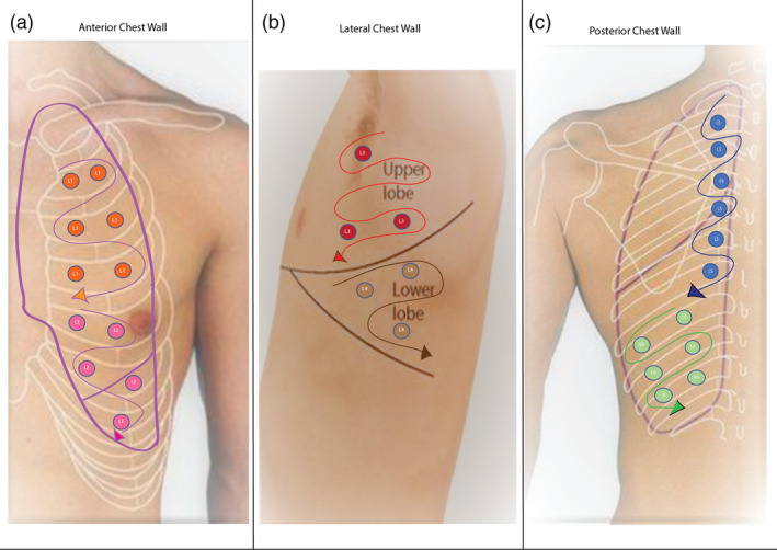
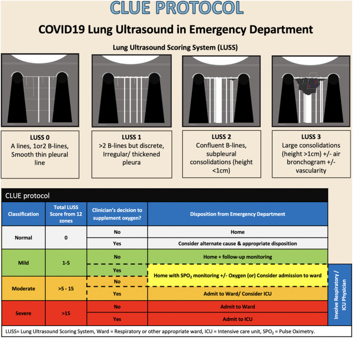
Lung ultrasound (LUS) plays a critical role in the SARS-CoV-2 pandemic. Evidence is mounting on its utility to diagnose, assess the severity and as a triage tool in the ED. Sonographic features correlate well to computed tomography (CT) chest findings and a bedside LUS performed by a trained clinician along with clinical examination, could be an alternative to chest X-ray and CT chest in these highly infectious patients. In this article, we have described a step-by-step approach to LUS in COVID patients and the CLUE (COVID-19 LUS in the ED) protocol, which involves an anatomical parameter, the severity of lung changes, objectively scored using the validated LUS scoring system and a physiological parameter, oxygen requirement. We believe this CLUE protocol can help risk-stratify patients presenting to ED with suspected COVID-19 and aid clinicians in making appropriate disposition decisions.
Keywords: CLUE; COVID-19; POCUS; emergency; lung ultrasound.
© 2020 Australasian College for Emergency Medicine.
Figures
References
-
- Mayo PH, Copetti R, Feller‐Kopman D et al. Thoracic ultrasonography: a narrative review. Intensive Care Med. 2019; 45: 1200–11. - PubMed
MeSH terms
LinkOut - more resources
Full Text Sources
Medical
Miscellaneous
