Case Reports
. 2020 May 10;14(1):57.
doi: 10.1186/s13256-020-02379-9.

Diffuse large B cell lymphoma involving Meckel's cave masquerading as biopsy-negative giant cell arteritis: a case report

Affiliations
Case Reports

Diffuse large B cell lymphoma involving Meckel's cave masquerading as biopsy-negative giant cell arteritis: a case report

Matthew J Samec et al. J Med Case Rep. .

Abstract

Background: Given the absence of consensus diagnostic criteria for giant cell arteritis, clinicians may encounter difficulty with identification of new-onset headache in patients older than age 50 years presenting with visual changes and elevated inflammatory markers, particularly if temporal artery biopsies are performed and negative.

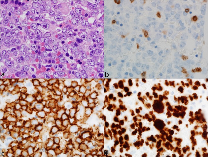
Case presentation: We present a case of a 57-year-old white man with headache, diplopia, and jaw paresthesia initially diagnosed and managed as steroid-refractory biopsy-negative giant cell arteritis. Further investigation disclosed evidence of soft tissue infiltration into Meckel's (trigeminal) cave bilaterally. Positron emission tomography suggested the presence of a lymphoproliferative disorder. Histology confirmed the diagnosis of diffuse large B cell lymphoma.

Conclusions: Metastatic involvement in Meckel's cave in diffuse large B cell lymphoma is extremely rare and presents a diagnostic challenge. Patients with suspicion of giant cell arteritis should undergo advanced imaging, particularly those with negative biopsy, atypical features, or lack of response to standard therapy, in order to assess for the presence of large-vessel vasculitis or other mimicking pathologies.

Keywords: Giant cell arteritis; Lymphoma; Metastatic; Trigeminal nerve.


Conflict of interest statement

The authors declare that they have no competing interests.

Figures

Fig. 1
Fig. 1
Diffuse large B cell lymphoma involvement on magnetic resonance imaging and positron emission tomography-computed tomography. Magnetic resonance images of brain show: a abnormal enhancement of Meckel’s cave bilaterally with extension through the foramen ovale (arrows) before chemotherapy and b decreased soft tissue in Meckel’s cave bilaterally (arrows) after chemotherapy. Positron emission tomography-computed tomography scans show: c extensive hypermetabolic lesions throughout the axial and appendicular skeleton, including the skull base, as well as fluorodeoxyglucose-avid lymph nodes above and below the diaphragm before chemotherapy and d marked response of all hypermetabolic lesions after chemotherapy
Fig. 2
Fig. 2
Bone marrow involvement by diffuse large B cell lymphoma. Photomicrographs of bone marrow trephine biopsy show: a medium to large neoplastic lymphocytes with nuclear irregularity, dispersed chromatin and variably sized nucleoli and background hematopoietic precursors on hematoxylin and eosin stain, b rare scattered T cells highlighted by CD3 immunohistochemistry, c membranous staining of neoplastic B cells by CD20 immunohistochemistry, and d nuclear staining of neoplastic B cells by PAX5 immunohistochemistry. a–d; × 1000
Fig. 3
Fig. 3
Algorithm for evaluation of suspected giant cell arteritis based on presentation features. CTA computed tomography angiography, GCA giant cell arteritis, MRA magnetic resonance angiography, MRI magnetic resonance imaging, PET-CT positron emission tomography-computed tomography, TA temporal artery, u/s ultrasound. (Adapted with permission from Koster MJ, Matteson EL, Warrington, KJ. Large-vessel giant cell arteritis: diagnosis, monitoring and management. Rheumatology 2018;57:ii32–ii42)

References

    1. Dasgupta B, Borg FA, Hassan N, Alexander L, Barraclough K, Bourke B, et al. BSR and BHPR guidelines for the management of giant cell arteritis. Rheumatology (Oxford) 2010;49(8):1594–1597. doi: 10.1093/rheumatology/keq039a. - DOI - PubMed
    1. Davies CG, May DJ. The role of temporal artery biopsies in giant cell arteritis. Ann R Coll Surg Engl. 2011;93(1):4–5. doi: 10.1308/003588411X12851639107476. - DOI - PMC - PubMed
    1. Koster MJ, Matteson EL, Warrington KJ. Large-vessel giant cell arteritis: diagnosis, monitoring and management. Rheumatology (Oxford) 2018;57(suppl_2):ii32–ii42. doi: 10.1093/rheumatology/kex424. - DOI - PubMed
    1. Donia MM, Gamaleldin OA, Abdo AM, Desouky SE, Helmy SAS. Intracranial neoplastic lesions of the trigeminal nerve: How MRI can help. Egypt J Radiol Nucl Med. 2017;48(4):1035–1041. doi: 10.1016/j.ejrnm.2017.07.008. - DOI
    1. Malhotra A, Tu L, Kalra VB, Wu X, Mian A, Mangla R, et al. Neuroimaging of Meckel's cave in normal and disease conditions. Insights Imaging. 2018;9(4):499–510. doi: 10.1007/s13244-018-0604-7. - DOI - PMC - PubMed

Publication types

MeSH terms

LinkOut - more resources