Review
. 2020 Jul;30(7):516-536.
doi: 10.1016/j.tcb.2020.04.005. Epub 2020 May 5.

Turning the Oxygen Dial: Balancing the Highs and Lows

Affiliations
Review

Turning the Oxygen Dial: Balancing the Highs and Lows

Alan H Baik et al. Trends Cell Biol. 2020 Jul.

Abstract

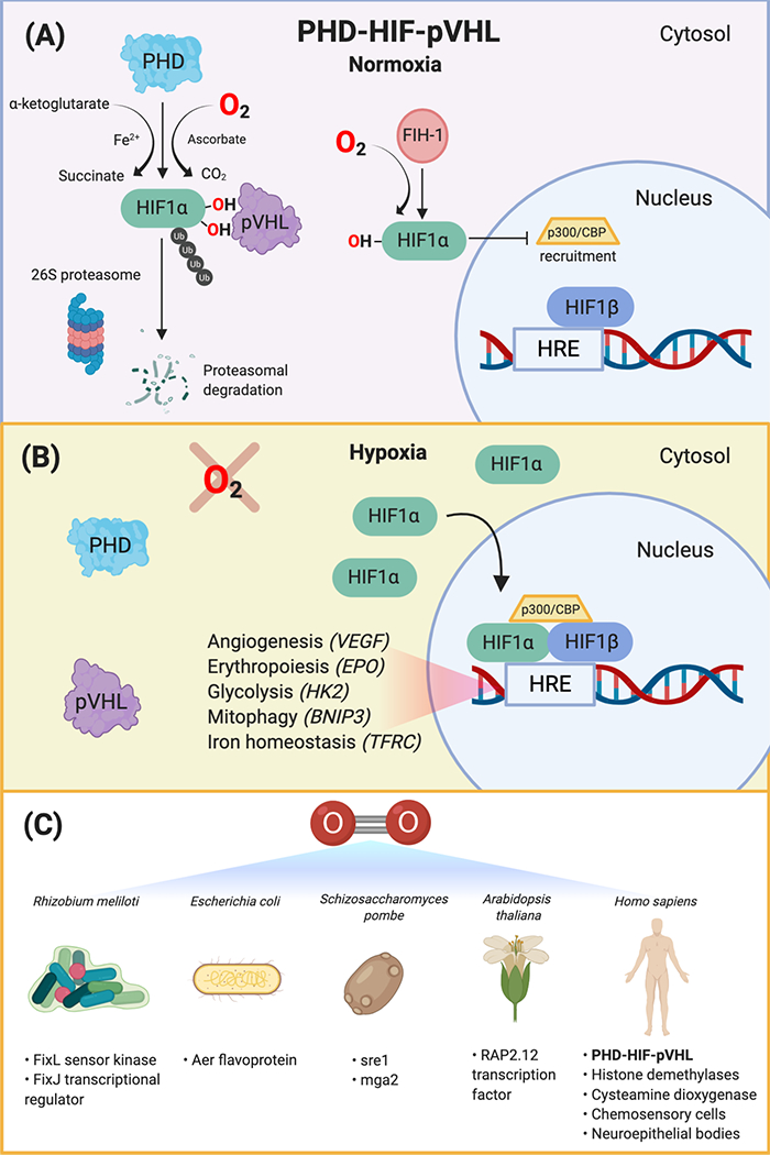
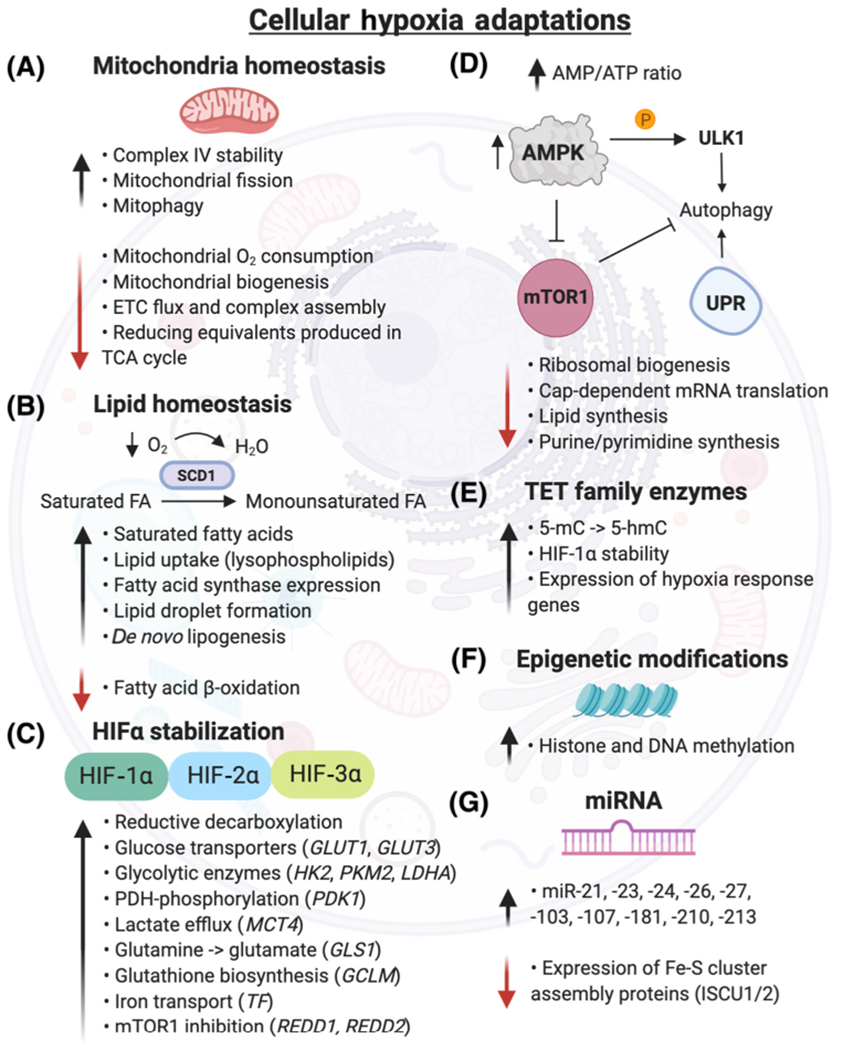
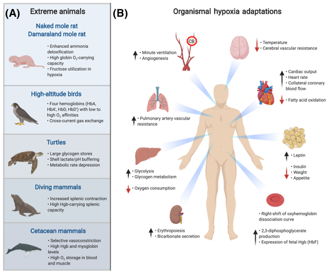
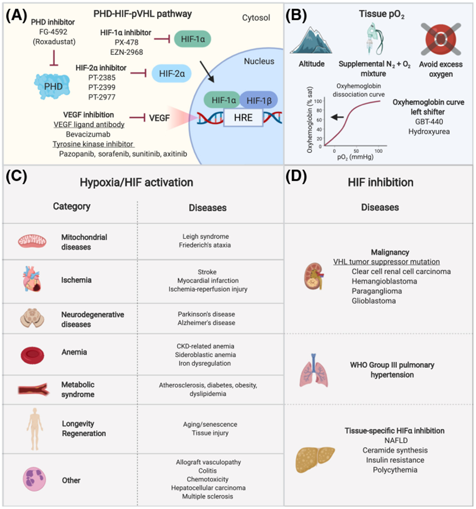
Oxygen is both vital and toxic to life. Molecular oxygen is the most used substrate in the human body and is required for several hundred diverse biochemical reactions. The discovery of the PHD-HIF-pVHL system revolutionized our fundamental understanding of oxygen sensing and cellular adaptations to hypoxia. It deepened our knowledge of the biochemical underpinnings of numerous diseases, ranging from anemia to cancer. Cellular dysfunction and tissue pathology can result from a mismatch of oxygen supply and demand. Recent work has shown that mitochondrial disease models display tissue hyperoxia and that disease pathology can be reversed by normalization of excess oxygen, suggesting that certain disease states can potentially be treated by modulating oxygen levels. In this review, we describe cellular and organismal mechanisms of oxygen sensing and adaptation. We provide a revitalized framework for understanding pathologies of too little or too much oxygen.

Keywords: hyperoxia; hypoxia; oxygen adaptation; oxygen metabolism; oxygen sensing.


Conflict of interest statement

Disclaimer Statement

A.H.B. has no conflicts of interest to declare. I.H.J. is listed on patents related to the use of hypoxia therapy for metabolic disorders.

Figures

Figure 1.
Figure 1.. Cellular Oxygen Sensors.
(A)The PHD-HIF-pVHL oxygen sensing pathway is conserved across all metazoans. Under normoxic conditions, PHD proteins hydroxylate the HIF-α transcription factor at two highly-conserved prolyl residues on the oxygen-dependent degradation domain, leading to its recognition by the tumor suppressor, pVHL. pVHL is the recognition component of a ubiquitin E3 ligase complex that polyubiquitylates HIF-α, marking it for proteasomal degradation by the 26S proteasome. Factor inhibiting HIF1 (FIH-1) is an asparagine hydroxylase that hydroxylates HIF in normoxia and prevents recruitment of the transcription coactivators, p300 and CBP. (B) Under hypoxic conditions, HIF-α is not degraded. HIF-α accumulates in the cytosol and translocates to the nucleus, where it binds to the conserved HRE sequence on DNA, its constitutively active and oxygen-insensitive partner, HIF-1β (ARNT), and the transcription coactivators, p300 and CBP. This complex transcriptionally activates hundreds of genes that allow cells to adapt to hypoxic environments, including VEGF, EPO, HK2, BNIP3, and TFRC. (C) There are various oxygen sensors across organisms, including Gram-negative bacteria (Rhizobium meliloti and Escherichia coli), fission yeast (Schizosaccharomyces pombe), plants (Arabidopsis thaliana), and metazoans (e.g., Homo sapiens). These systems, including the FixL sensor kinase and FixJ transcriptional response regulator, Aer flavoprotein, sre1 and mga2 transcriptional regulators, RAP2.12 transcription factor, histone demethylases, chemosensory cells in the carotid body, and intrapulmonary neuroepithelial bodies, transduce oxygen-dependent reactions into a signaling cascade that result in adaptive responses in hypoxic conditions. Abbreviations: BNIP3, BCL2 interacting protein 3; CBP, cyclic-AMP response element binding protein (CREB) binding protein; EPO, erythropoietin; TFRC, transferrin receptor; HIF, hypoxia-inducible factor; HK2, hexokinase-2, HRE, hypoxia response element; PHD, prolyl hydroxylase domain; pVHL, von-Hippel-Lindau tumor suppressor protein; VEGF, vascular endothelial growth factor.
Figure 2.
Figure 2.. Cellular Hypoxia Adaptations.
(A) Mitochondria adapt to hypoxia by increasing cytochrome c oxidase (complex IV) stability, which facilitates electron transport to O2. Mitochondria biogenesis, ETC flux, and respiration are downregulated, reducing O2 consumption and reactive oxygen species production [223]. Hypoxia induces mitochondrial fission by increasing the activity of the E3 ubiquitin ligase, SIAH2, which degrades AKAP121, subsequently increasing Drp1/Fis1 interaction [224]. (B) Under hypoxia, cells become deficient in unsaturated fatty acids by reduced O2-dependent desaturation of saturated fatty acids by stearoyl-CoA desaturases (e.g., SCD1). Hypoxic cells increase uptake of lysophospholipids and formation of lipid droplets, which serve as extra supply stores for lipids and help buffer against saturated lipid species [225]. Hypoxia downregulates fatty acid β-oxidation [226]. (C) Gene expression of several HIF targets are highlighted, though hundreds of genes are regulated by HIF stabilization [24]. (D) Hypoxia andthe subsequent energy deficiency (elevated AMP/ATP ratio) inhibit mTOR1 through AMP-activated protein kinase (AMPK). The downstream pathways regulated by mTOR1, including ribosomal and lipid synthesis, are downregulated in hypoxia, while apoptosis is increased. Unc-51-like kinase 1 (ULK1), an autophagy initiation serine/threonine protein kinase, is directly phosphorylated by AMPK in hypoxia, initiating a proautophagy signal [227]. Severe hypoxia (<0.01% O2) promotes the transcriptional response of essential autophagy genes and activates the UPR in a HIF-independent manner [228]. (E) Ten-eleven translocation methylcytosine dioxygenases (TET) catalyze the conversion of 5-methylcytosine (5-mC) to 5-hydroxymethylcytosine (5-hmC), increasing DNA methylation and gene expression of hypoxia responsive genes [8]. (F) Hypoxia induces changes in histone and DNA methylation via KDM and TET enzymes, respectively, which regulate chromatin and methylation homeostasis. (G) Numerous microRNAs are induced by hypoxia. miR-210 is increased in a HIF-1α-dependent manner and represses iron-sulfur cluster assembly proteins (ISCU1/2), resulting in decreased integrity of Fe-S cluster proteins [53]. Abbreviations: ETC, electron transport chain; FA, fatty acid; GCLM, glutamate-cysteine ligase modifier subunit; GLS1, glutaminase; GLUT, glucose transporter; HIF, hypoxia-inducible factor; HK2, hexokinase 2; KDM, lysine demethylase; LDHA, lactate dehydrogenase A; MCT4, monocarboxylate transporter 4; mTOR, mammalian target of rapamycin; PDK1, pyruvate dehydrogenase kinase; PKM2, pyruvate kinase muscle isozyme 2; REDD1/2, DNA damage response; TCA, tricarboxylic acid cycle; TF, transferrin; UPR, unfolded protein response.
Figure 3.
Figure 3.. Organismal Hypoxia Adaptations in Extreme Animals and Homo sapiens.
(A) The naked mole rat (Heterocephalus glaber), high-altitude birds (e.g., Gyps rueppelli), turtles (e.g., Chrysemys picta), diving mammals (e.g., Leptonychotes weddellii), and cetacean mammals (e.g., Tursiops truncatus) [229], have developed unique metabolic and physiologic adaptations to extreme hypoxic environments. (B) Hypoxia induces whole-body physiologic and tissue metabolic adaptations in Homo sapiens involving the brain, heart, adipose tissue, kidney, liver, pulmonary and systemic vasculature, and carotid body (CB). Hypoxia induces generalized vasodilation with the exception of HIF-2α-mediated vasoconstriction of pulmonary arteries, leading to pulmonary hypertension. Metabolic changes include increased circulating leptin levels, decreased insulin levels, and increased glycogen metabolism [230]. Cardiac adaptations include increased heart rate, cardiac output, and coronary artery blood flow, and decreased fatty acid oxidation rate [231]. Chemosensory type I glomus cells in the CB sense hypoxia and increase the ventilation rate via activation of action potentials in the glossopharyngeal nerve that excite central chemoreceptors in the brain. Kidneys adapt by increasing bicarbonate secretion and erythropoiesis via HIF-2α-dependent induction of erythropoietin, augmenting oxygen-carrying capacity to tissues. The oxyhemoglobin dissociation curve shifts to the right, facilitating unloading of oxygen into tissues. Hypoxia promotes erythroid expansion of mature red blood cell (RBC) precursors containing fetal hemoglobin (HbF) [232]. Abbreviation: Hgb, hemoglobin.
Figure 4.
Figure 4.. Hypoxia Response Therapy and Candidate Diseases Amenable to Hypoxia/HIF Activation or HIF Inhibition.
(A) Medications and novel small-molecule compounds that inhibit prolyl hydroxylase domain (PHD) proteins, HIF-1α, HIF-2α, and VEGF have shown clinical benefit in preclinical models and clinical trials [153,154,183,185,233]. PHD inhibition increases the HIF transcriptional response by preventing HIF hydroxylation, whereas HIF-1α and HIF-2α inhibition downregulate the HIF response. (B) Hypobaric hypoxia can be achieved by ascent to altitude or use of hypobaric hypoxia chambers, which simulate hypoxia by reducing atmospheric pressure. Normobaric hypoxia can be achieved by delivery of a mixture of nitrogen and oxygen gases and pressure swing adsorption systems. Tissue hypoxia can be mimicked by administration of oxyhemoglobin curve left shifters. Certain mitochondrial diseases are associated with tissue hyperoxia and preclinical studies show that pathologic phenotypes can be ameliorated by hypoxia exposure. In these conditions, excess oxygen should be avoided unless clinically indicated. (C) Category and examples of diseases that can potentially be treated with hypoxia or HIF activation (e.g., PHD inhibition). (D) Candidate diseases that can be treated with inhibition of the HIF response (e.g., HIF or VEGF inhibition). Abbreviations: CKD, chronic kidney disease; HIF, hypoxia-inducible factor; HRE, hypoxia response element; NAFLD, non-alcoholic fatty liver disease; PHD, prolyl hydroxylase domain protein; pO2, oxygen tension; VEGF, vascular endothelial growth factor; VHL, von Hippel-Lindau tumor suppressor protein; WHO, World Health Organization.

References

    1. Gumsley AP et al. (2017) Timing and tempo of the Great Oxidation Event. Proc. Natl. Acad. Sci. U. S. A 114, 1811–1816 - PMC - PubMed
    1. Severinghaus JW (2003) Fire-air and dephlogistication. Revisionisms of oxygen’s discovery. Adv. Exp. Med. Biol 543, 7–19 - PubMed
    1. Ashcroft FM (2000) Life at the Extremes, University of California Press
    1. Dejours P and Dejours S (1992) The effects of barometric pressure according to Paul Bert: the question today. Int. J. Sports Med 13, S1S–5 - PubMed
    1. Carreau A et al. (2011) Why is the partial oxygen pressure of human tissues a crucial parameter? Small molecules and hypoxia. J. Cell. Mol. Med 15, 1239–1253 - PMC - PubMed

Publication types

Substances