

Case Reports
doi: 10.1016/j.thromres.2020.04.046.
Epub 2020 May 1.
Arterial and venous thromboembolic disease in a patient with COVID-19: A case report
Affiliations
- PMID: 32386986
- PMCID: PMC7252130
- DOI: 10.1016/j.thromres.2020.04.046
Item in Clipboard
Case Reports
Arterial and venous thromboembolic disease in a patient with COVID-19: A case report
Thromb Res.
2020 Jul.
No abstract available
Conflict of interest statement
Declaration of competing interest The authors have declared no conflicts of interest.
Figures

CT scans of the chest. (A) High resolution chest CT scan performed at admission, showing ground-glass abnormalities with and without reticulation (“crazy paving”) with a predominantly peripheral distribution, consistent with COVID-19-related pneumonia. (B) CTPA performed at admission that did not reveal signs of pulmonary emboli. (C) High resolution chest CT scan performed on day 2, showing a marked increase in COVID-19-related pulmonary involvement and new areas of consolidation. (D) CTPA performed on day 2, again showing no signs of pulmonary embolism. (E) High resolution chest CT scan performed on day 7, showing improvement of COVID-19-related pulmonary changes with less extensive abnormalities and signs compatible with organising pneumonia. (F) CTPA performed on day 7, showing lobar (arrow) and subsegmental (not shown) pulmonary emboli.
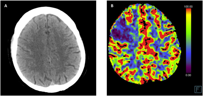
CT imaging of the brain. (A) CT excluding brain haemorrhage. (B) CT-perfusion showing a defect right frontal with large mismatch between perfusion and vascular volume recordings, indicative of right frontal lobe infarction.
Dataset described in
-
Incidence of thrombotic complications in critically ill ICU patients with COVID-19.Thromb Res. 2020 Jul;191:145-147. doi: 10.1016/j.thromres.2020.04.013. Epub 2020 Apr 10. Thromb Res. 2020. PMID: 32291094 Free PMC article.
References
-
- Ekker M.S., Boot E.M., Singhal A.B., Tan K.S., Debette S., Tuladhar A.M., et al. Epidemiology, aetiology, and management of ischaemic stroke in young adults. Lancet Neurol. 2018;17:790–801. - PubMed
Publication types
MeSH terms
Substances
LinkOut - more resources
Full Text Sources
Other Literature Sources
Medical
