Review
. 2020 Sep;17(9):1463-1471.
doi: 10.1016/j.hrthm.2020.05.001. Epub 2020 May 5.

Recognizing COVID-19-related myocarditis: The possible pathophysiology and proposed guideline for diagnosis and management

Affiliations
Review

Recognizing COVID-19-related myocarditis: The possible pathophysiology and proposed guideline for diagnosis and management

Bhurint Siripanthong et al. Heart Rhythm. 2020 Sep.

Abstract

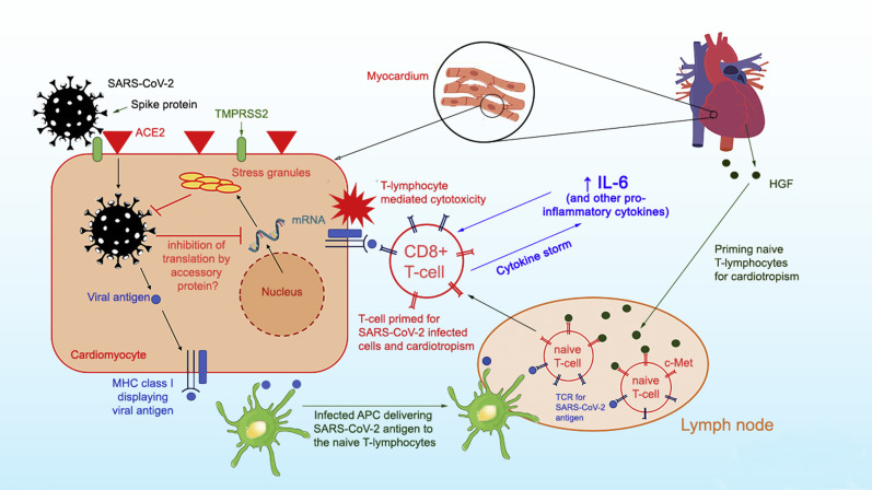
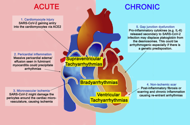
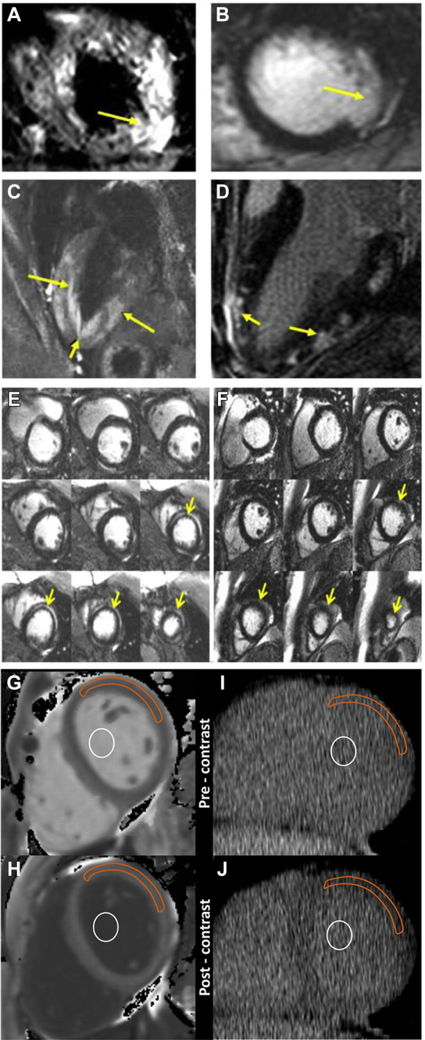
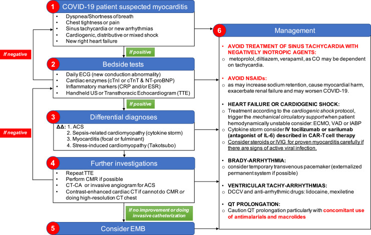
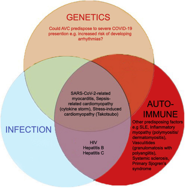
Human coronavirus-associated myocarditis is known, and a number of coronavirus disease 19 (COVID-19)-related myocarditis cases have been reported. The pathophysiology of COVID-19-related myocarditis is thought to be a combination of direct viral injury and cardiac damage due to the host's immune response. COVID-19 myocarditis diagnosis should be guided by insights from previous coronavirus and other myocarditis experience. The clinical findings include changes in electrocardiogram and cardiac biomarkers, and impaired cardiac function. When cardiac magnetic resonance imaging is not feasible, cardiac computed tomographic angiography with delayed myocardial imaging may serve to exclude significant coronary artery disease and identify myocardial inflammatory patterns. Because many COVID-19 patients have cardiovascular comorbidities, myocardial infarction should be considered. If the diagnosis remains uncertain, an endomyocardial biopsy may help identify active cardiac infection through viral genome amplification and possibly refine the treatment risks of systemic immunosuppression. Arrhythmias are not uncommon in COVID-19 patients, but the pathophysiology is still speculative. Nevertheless, clinicians should be vigilant to provide prompt monitoring and treatment. The long-term impact of COVID-19 myocarditis, including the majority of mild cases, remains unknown.

Keywords: Arrhythmias; Coronavirus disease 2019; Endomyocardial biopsy; Fulminant myocarditis; Interleukin 6; SARS-CoV-2.


Figures

Figure 1
Figure 1
Proposed pathophysiology of SARS-CoV-2 myocarditis. SARS-CoV-2 utilizes the spike protein (primed by TMPRSS2) to bind ACE2 to allow cell entry. Intracellular SARS-CoV-2 might impair stress granule formation via its accessory protein. Without the stress granules, the virus is allowed to replicate and damage the cell. Naïve T lymphocytes can be primed for viral antigens via antigen-presenting cells and cardiotropism by the heart-produced HGF. The HGF binds c-Met, an HGF receptor on T lymphocytes. The primed CD8+ T lymphocytes migrate to the cardiomyocytes and cause myocardial inflammation through cell-mediated cytotoxicity. In the cytokine storm syndrome, in which proinflammatory cytokines are released into the circulation, T-lymphocyte activation is augmented and releases more cytokines. This results in a positive feedback loop of immune activation and myocardial damage. ACE2 = angiotensin-converting enzyme 2; APC = antigen-presenting cell; HGF = hepatocyte growth factor; IL-6 = interleukin 6; MHC = major histocompatibility complex; SARS-CoV-2 = severe acute respiratory syndrome coronavirus 2; TCR = T-cell receptor.
Figure 2
Figure 2
Arrhythmogenesis in SARS-CoV-2–related myocarditis. Possible mechanisms responsible for arrhythmias in SARS-CoV-2–related myocarditis are shown. Mechanisms 1, 2, and 3 could occur in the acute setting, whereas mechanisms 4 and 5 occur in chronic/healed myocarditis. Abbreviations as in Figure 1.
Figure 3
Figure 3
Typical cardiovascular magnetic resonance (CMR) and computed tomography (CT) findings of myocarditis. A, C: Cardiac edema (yellow arrows) in T2-weighted mode. B, D: LGE of the subepicardial region (yellow arrows) of the ventricles, a sign of myocardial fibrosis or scarring. E: Midmyocardial LGE (yellow arrows) is often present in the acute setting and resolved at follow-up. F: LGE resolved in the chronic case. These areas may initially represent acute myocardial edema (yellow arrows) and resolve over time. G, H: CMR imaging of region-of-interest measurements obtained before (G) and after (H) gadolinium chelate administration. I, J: Reformatted cardiac CT of region-of-interest measurements obtained before (I) and after (J) administration of an iodinated contrast agent. For cardiac CT, the anterolateral myocardium was most reliably identified before administration of an iodinated contrast agent. In that area, a region of interest from the anterolateral myocardium was used for attenuation measurements. A focal myocardial scar was identified on delayed CMR images and was not included in the region of interest. Orange outline indicates myocardium; white circle indicates blood pool. LGE = late gadolinium enhancement. A-F courtesy of Dr Raymond Y. Kwong (Brigham and Women's Hospital), G-J reprinted from Nacif et al, Radiology, 2012, vol 264, p.876-883, with permission from RSNA.
Figure 4
Figure 4
Suggested diagnostic and management protocol for SARS-CoV-2–related myocarditis. ΔΔ = differential diagnoses; ACS = acute coronary syndrome; CAR = chimeric antigen receptor; CMR = cardiovascular magnetic resonance; CO = cardiac output; COVID-19 = coronavirus 19; CRP = C-reactive protein; CT = computed tomography; CT-CA = computed tomography–coronary angiogram; cTnI = cardiac troponin I; cTnT = cardiac troponin T; DCCV = direct current cardioversion; ECG = electrocardiogram; ECMO = extracorporeal membrane oxygenation; EMB = endomyocardial biopsy; ESR = erythrocyte sedimentation rate; IABP = intra-aortic balloon pump; IL-6 = interleukin 6; IV = intravenous; IVIG = intravenous immunoglobulin; NSAID = nonsteroidal anti-inflammatory drug; NT-proBNP = N-terminal pro–B-type natriuretic peptide; SARS-CoV-2 = severe acute respiratory syndrome coronavirus 2; TTE = transthoracic echocardiogram; US = ultrasound; VAD = ventricular assist device.
Supplementary Figure 1

References

    1. Zhu N., Zhang D., Wang W. A novel coronavirus from patients with pneumonia in China, 2019. N Engl J Med. 2020;382:727–733. - PMC - PubMed
    1. Mahase E. Covid-19: death rate is 0.66% and increases with age, study estimates. BMJ. 2020;369:m1327. - PubMed
    1. Zeng J.-H., Liu Y.-X., Yuan J. First case of COVID-19 infection with fulminant myocarditis complication: case report and insights. Published online April 10. Infection. 2020 doi: 10.1007/s15010-020-01424-5. - DOI - PMC - PubMed
    1. Inciardi R.M., Lupi L., Zaccone G. Cardiac involvement in a patient with coronavirus disease 2019 (COVID-19) JAMA Cardiol. 2020;5:819–824. - PMC - PubMed
    1. Kim I.-C., Kim J.Y., Kim H.A., Han S. COVID-19-related myocarditis in a 21-year-old female patient. Eur Heart J. 2020;41:1859. - PMC - PubMed