Review
. 2020 Oct-Dec;26(5-6):503-520.
doi: 10.1177/1073858420916696. Epub 2020 May 9.

The Accumulation of Tau in Postsynaptic Structures: A Common Feature in Multiple Neurodegenerative Diseases?

Affiliations
Review

The Accumulation of Tau in Postsynaptic Structures: A Common Feature in Multiple Neurodegenerative Diseases?

Peter J Teravskis et al. Neuroscientist. 2020 Oct-Dec.

Abstract

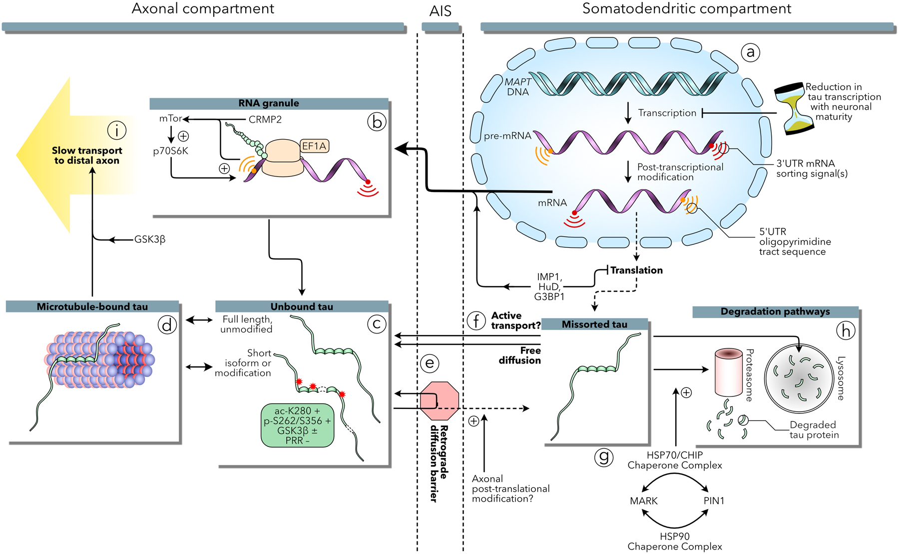
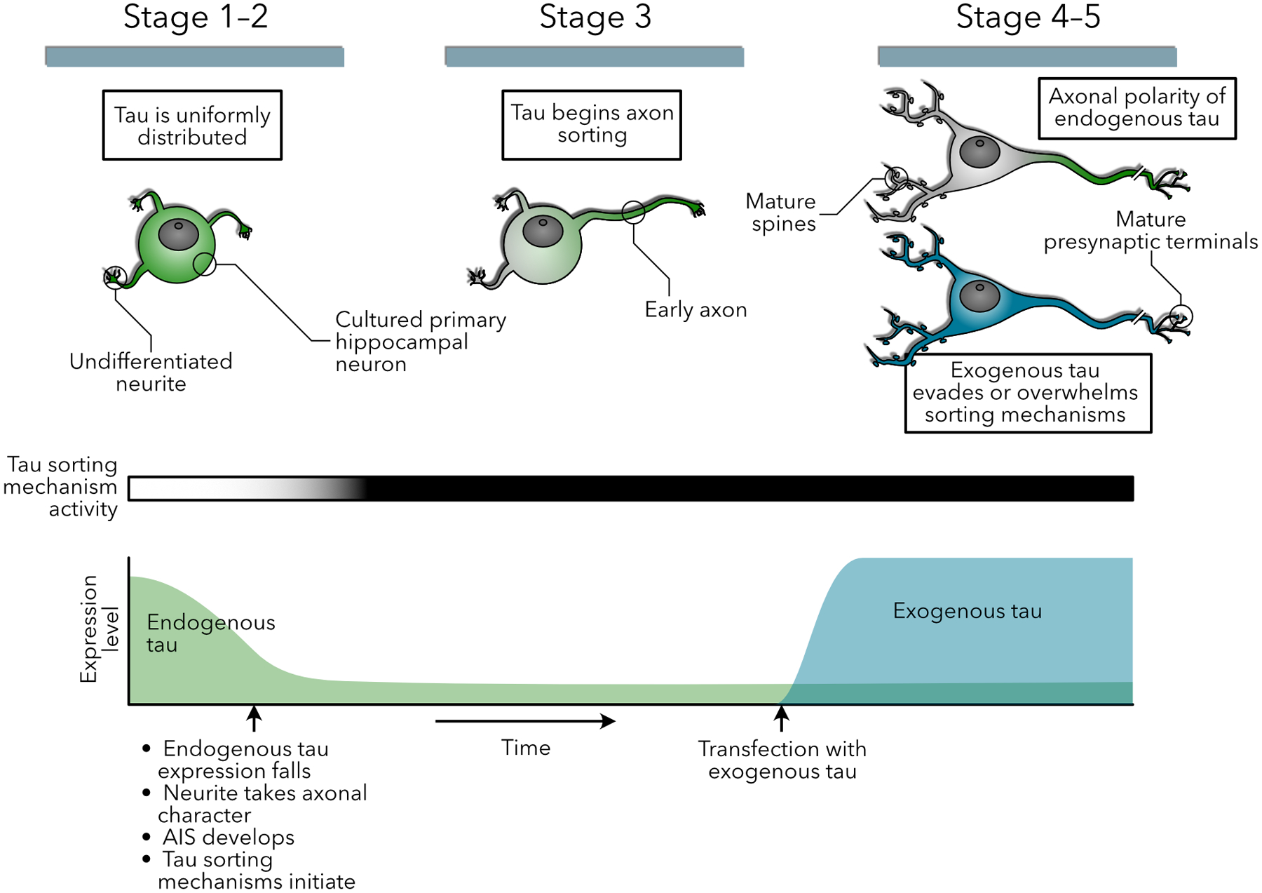
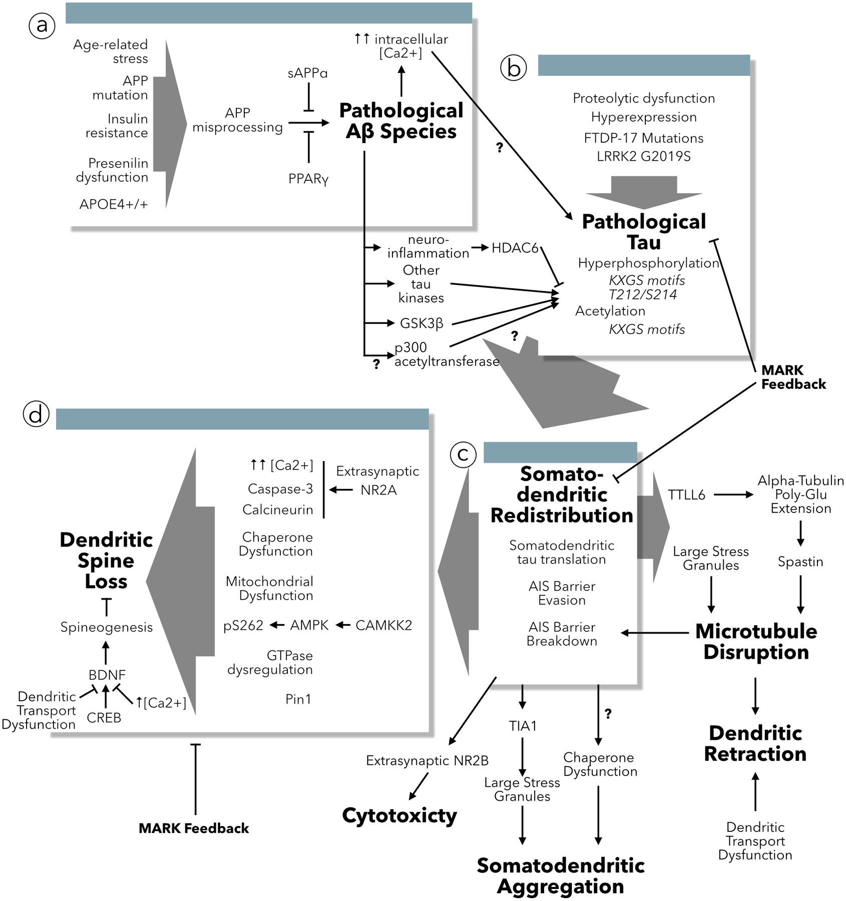
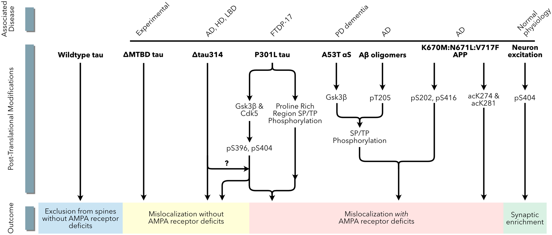
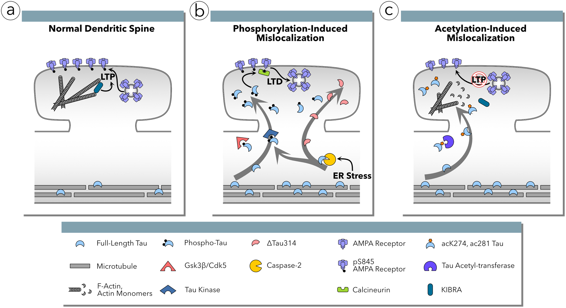
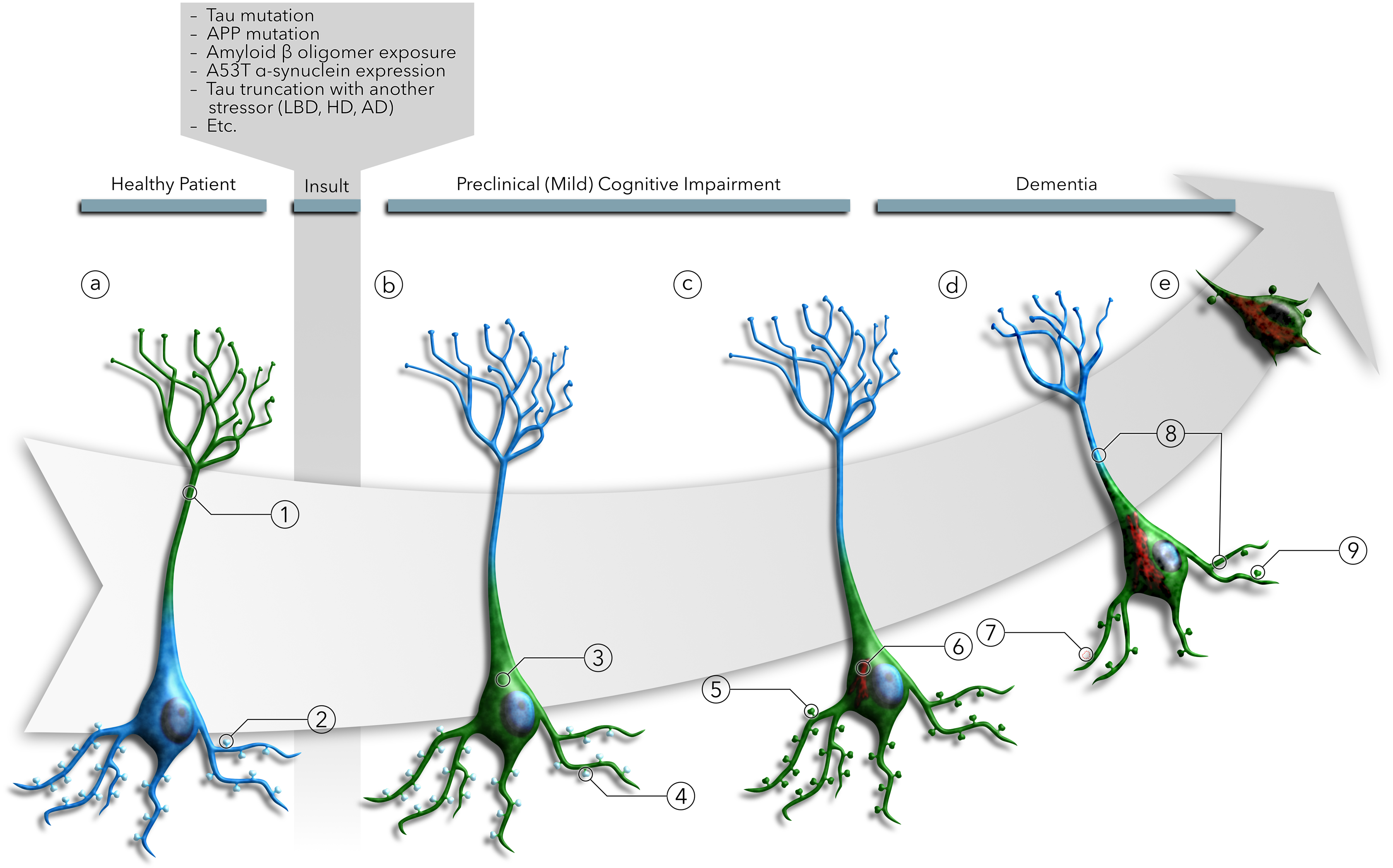
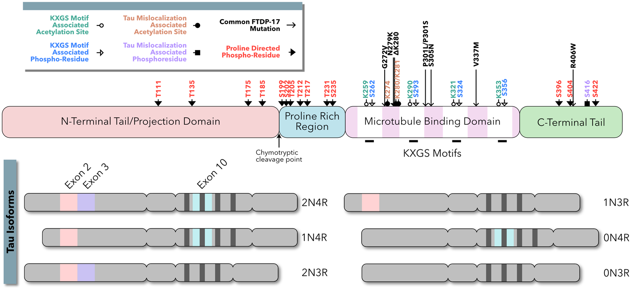
Increasingly, research suggests that neurodegenerative diseases and dementias are caused not by unique, solitary cellular mechanisms, but by multiple contributory mechanisms manifesting as heterogeneous clinical presentations. However, diverse neurodegenerative diseases also share common pathological hallmarks and cellular mechanisms. One such mechanism involves the redistribution of the microtubule associated protein tau from the axon into the somatodendritic compartment of neurons, followed by the mislocalization of tau into dendritic spines, resulting in postsynaptic functional deficits. Here we review various signaling pathways that trigger the redistribution of tau to the cell body and dendritic tree, and its mislocalization to dendritic spines. The convergence of multiple pathways in different disease models onto this final common pathway suggests that it may be an attractive pathway to target for developing new treatments for neurodegenerative diseases.

Keywords: AMPA receptors; Alzheimer’s disease; FTDP-17; Huntington’s disease; MAPT; Parkinson’s disease; dendritic spines; neuronal polarity; synaptic plasticity; tau.


Conflict of interest statement

CONFLICTS: None

Figures

Figure 1:
Figure 1:
Mechanisms underlying the axonal localization of tau in healthy neurons. (a) The MAPT gene codes for 5’ and 3’UTR signals critical to tau mRNA sorting and selective translation in the axon. After a short burst of expression early in development, tau mRNA transcription is significantly reduced with neuronal maturation by an unknown antenatal mechanism. Tau mRNA may be preserved by the contribution of IMP1, HuD, and G3BP1 during transit to the axon and those proteins may contribute to suppressing expression in the somatodendritic compartment. (b) Tau mRNA undergoes active translation in RNA granules in the axon. The 5’UTR oligopyramidal tract signals of tau and CRMP2 mRNA encourage selective translation via an mTor-p70S6K mechanism. (c) Once translated, tau protein can bind axonal microtubules (d), tau isoform and posttranslational modifications have been shown to have differential effects on microtubule binding that may contribute to efflux from the axon through a retrograde diffusion barrier associated with the axon initial segment (AIS) (e). Tau translated in the somatodendritc compartment can freely diffuse into the axon, or may be involved in an unknown active transport mechanism (f). Alternatively, somatodenritic tau is degraded by proteasome and lysosome proteolytic mechanisms, potentially regulated by the interplay of MARK and Pin1 within a chaperone protein refolding pathway (g–h). Finally, tau may progress into the distal axon via slow transport on microtubule segments or by kinesin motor transport, regulated by GSK3β (i).
Figure 2:
Figure 2:
Hypothetical model of the temporal relationship between sorting mechanisms and endogenous and exogenous tau expression. (Top row) Diagrams of endogenous tau (green) and exogenous tau (blue) distribution in a developing cultured hippocampal neuron. While tau is initially uniformly distributed throughout the cellular sphereoid and early, undifferentiated neuronal processes, sorting begins once one of the processes takes on axonal character, and is complete with the elaboration of mature postsynaptic morphology. This is dependent on two mechanisms (1) a drop in mRNA levels and new tau expression shortly after birth, and (2) the initiation of tau sorting mechanisms upon development of axonal characteristics. However, the high expression levels of exogenous tau overcome these sorting mechanisms, especially in the absence of mRNA sorting signals.
Figure 3:
Figure 3:
Causes and consequences of tau redistribution to dendrites. (a) Multiple triggers encourage the misprocessing of APP leading to the accumulation of pathogenic Aβ species. Aβ increases intracellular calcium concentration and acts through multiple mediators to change the post-translational modification state of tau. (b) Aside from Aβ, multiple mechanisms can induce changes in tau that lead missorting to dendrites. (c) Once tau has been redistributed into dendrites it effectuates multiple pathologies including disrupting microtubules leading to dendritic retraction and transport dysfunction, undergoing misfolding and aggregation, and mediating cytotoxicity. (d) The primary result of tau redistribution to dendrites is dendritic spine loss, which has been associated with multiple protein signaling cascades as well as potential disruption of BDNF spine stabilization and spinogenesis. This may or may not be associated with tau-induced dendritic microtubule transport dysfunction.
Figure 4:
Figure 4:
Multiple pathways in multiple disease models lead to tau mislocalization to dendritic spines. Models of AD, HD, PDD, FTDP-17 and LBD all implicate tau mislocalization to dendritic spines via phosphorylation or acetylation posttranslational modifications. The phosphorylation pattern of tau leading to mislocalization may be different in each condition.
Figure 5:
Figure 5:
Tau mislocalization to dendritic spines and AMPA receptor functional deficits are mediated by sequential phosphorylation in two domains and acetylation posttranslational modifications. (a) Under normal conditions, tau is excluded from the dendritic spines. The AMPA receptor subunit GluA1 is phosphorylated at Ser845 and the population of AMPA receptors in the postsynaptic membrane is stable. F-actin polymerization supports KIBRA-mediated, activity-induced insertion of AMPA receptors into the postsynaptic membrane, promoting memory formation via long-term potentiation (LTP). (b) In FTDP-17, AD, LBD, PDD and HD, tau is phosphorylated by CDK5 or GSK3β in the C-terminal domain at Ser396 and Ser404 residues resulting in mislocalization to dendritic spines. Mislocalization alone is insufficient to result in functional deficits; separate phosphorylation in the proline rich region (PRR) activates a signaling cascade in which calcineurin dephosphorylates GluA1 at Ser845 leading to AMPA receptor internalization and deficits in excitatory synaptic transmission resembling long-term depression (LTD). This pathway requires the activity of caspase-2 resulting the abnormal cleavage of tau at Asp314 creating Δtau314, which mislocalizes to dendritic spines but cannot induce synaptic deficits without some other cellular stressor. (c) In human AD, tau is abnormally acetylated at residues Lys274 and Lsy281 by an unknown acetyltransferase. Acetylation at these sites results in mislocalization to dendritic spines, actin depolymerization, and reduced KIBRA activity resulting in decreased AMPA receptor insertion into the postsynaptic membrane during LTP induction.
Figure 6:
Figure 6:
Illustration of the tau missorting sequence. (a) In healthy neurons, tau (green) is (a1) localized to the axon at levels three times greater than the soma and dendrites. (a2) While tau is largely excluded from dendritic spines, the small amount of tau located in the soma and dendrites may enter the spines and facilitate synaptic strengthening during LTP induction. After pathogenic insults from multiple disease mechanisms, (b3) tau is redistributed into the soma and dendrites of neurons due to the breakdown of tau sorting mechanisms or tau modifications leading to evasion of those mechanisms. However, (b4) dendritic tau does not mislocalize to dendritic spines. Once in the dendritic shaft, (c5) phosphorylation, truncation, and acetylation of tau lead to further mislocalization into the dendritic spines. (c6) Tau, redistributed in the soma and dendrites, begins to aggregate to form tau inclusions. Somatodendritic tau redistribution leads to (d7) dendritic spine loss, and (d8) retraction of neuritic processes. (d9) Tau mislocalization to dendritic spines leads to AMPA receptor signaling deficits due to LTD-like AMPA receptor internalization and the failure of LTP induction mechanisms. (e) Ultimately, somatodendritic tau contributes to cytotoxicity and cell death. Notably, overt tau pathology and neuron loss have been shown to be dissociated, even in the presence of pro-apoptotic signals, suggesting that cell death may result from the contribution of an independent pathway.
None

References

    1. Amar F, Sherman M, Rush T, Larson M, Boyle G, Chang L. and et al.. 2017. The amyloid-β oligomer Aβ*56 induces specific alterations in neuronal signaling that lead to tau phosphorylation and aggregation. Sci Signal. 10:1–14. - PMC - PubMed
    1. Aronov S, Aranda G, Behar L, and Ginzburg I. 2001. Axonal tau mRNA localization coincides with tau protein in living neuronal cells and depends on axonal targeting signal. J Neurosci. 21(17):6577–87. - PMC - PubMed
    1. Asthana J, Kapoor S, Mohan R, and Panda D. 2013. Inhibition of HDAC6 deacetylase activity increases its binding with microtubules and suppresses microtubule dynamic instability in MCF-7 cells. J Biol Chem. 288(31):22516–26. - PMC - PubMed
    1. Atlas R, Behar L, Sapoznik S, and Ginzburg I. 2007. Dynamic association with polysomes during P19 neuronal differentiation and an untranslated-region-dependent translation regulation of the tau mRNA by the tau mRNA-associated proteins IMP1, HuD, and G3BP1. J Neurosci Res. 85(1):173–83. - PubMed
    1. Balaji V, Kaniyappan S, Mandelkow E, Wang Y, and Mandelkow E. 2018. Pathological missorting of endogenous MAPT/Tau in neurons caused by failure of protein degradation systems. Autophagy. 14(12):2169–54. - PMC - PubMed

Publication types