

Bimodal magnetic resonance and optical imaging of extracellular matrix remodelling by orthotopic ovarian tumours
- PMID: 32390007
- PMCID: PMC7374547
- DOI: 10.1038/s41416-020-0878-7
Bimodal magnetic resonance and optical imaging of extracellular matrix remodelling by orthotopic ovarian tumours
Abstract
Background: The extracellular matrix modulates the development of ovarian tumours. Currently, evaluation of the extracellular matrix in the ovary is limited to histological methods. Both magnetic resonance imaging (MRI) and two-photon microscopy (2PM) enable dynamic visualisation and quantification of fibrosis by endogenous contrast mechanisms: magnetisation transfer (MT) MRI and second-harmonic generation (SHG) 2PM, respectively.
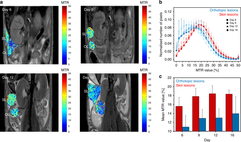
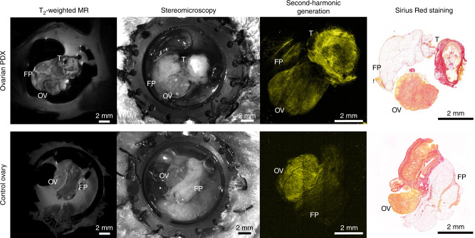
Methods: Here, we applied the MT-MRI protocol for longitudinal imaging of the stroma in orthotopic human ovarian cancer ES-2 xenograft model in CD1 athymic nude mice, and for orthotopically implanted ovarian PDX using a MR-compatible imaging window chamber implanted into NSG mice.
Results: We observed differences between ECM deposition in ovarian and skin lesions, and heterogeneous collagen distribution in ES-2 lesions. An MR-compatible imaging window chamber enabled visual matching between T2 MRI maps of orthotopically implanted PDX grafts and anatomical images of their microenvironment acquired with a stereomicroscope and SHG-2PM intravital microscopy of the collagen. Bimodal MRI/2PM imaging allowed us to quantify the fibrosis within the same compartments, and demonstrated the consistent results across the modalities.
Conclusions: This work demonstrates a novel approach for measuring the stromal biomarkers in orthotopic ovarian tumours in mice, on both macroscopic and microscopic levels.
Conflict of interest statement
The authors declare no competing interests.
Figures


References
-
- Siegel RL, Miller KD, Jemal AJCacjfc. Cancer statistics, 2017. CA: Cancer J. Clin. 2017;67:7–30. - PubMed
-
- Davis A, Tinker AV, Friedlander MJGo. “Platinum resistant” ovarian cancer: what is it, who to treat and how to measure benefit? Gynecol. Oncol. 2014;133:624–631. - PubMed
-
- Helleman J, Jansen MP, Burger C, van der Burg ME, Berns EMJTijob, biology c. Integrated genomics of chemotherapy resistant ovarian cancer: a role for extracellular matrix, TGFbeta and regulating microRNAs. Int. J. Biochem. Cell Biol. 2010;42:25–30. - PubMed
-
- Zhang Y, Tang H, Cai J, Zhang T, Guo J, Feng D, et al. Ovarian cancer-associated fibroblasts contribute to epithelial ovarian carcinoma metastasis by promoting angiogenesis, lymphangiogenesis and tumor cell invasion. Cancer Lett. 2011;303:47–55. - PubMed
Publication types
MeSH terms
Substances
Grants and funding
- R01 CA075334/CA/NCI NIH HHS/United States
- 326/14/Israel Science Foundation (ISF)
- 232640-IMAGO/EC | EC Seventh Framework Programm | FP7 Ideas: European Research Council (FP7-IDEAS-ERC - Specific Programme: "Ideas" Implementing the Seventh Framework Programme of the European Community for Research, Technological Development and Demonstration Activities (2007 to 2013))
LinkOut - more resources
Full Text Sources
Medical
