Review
. 2020 Jun 8;49(11):3278-3296.
doi: 10.1039/d0cs00181c.

Surface analysis tools for characterizing biological materials

Affiliations
Review

Surface analysis tools for characterizing biological materials

Joe E Baio et al. Chem Soc Rev. .

Abstract

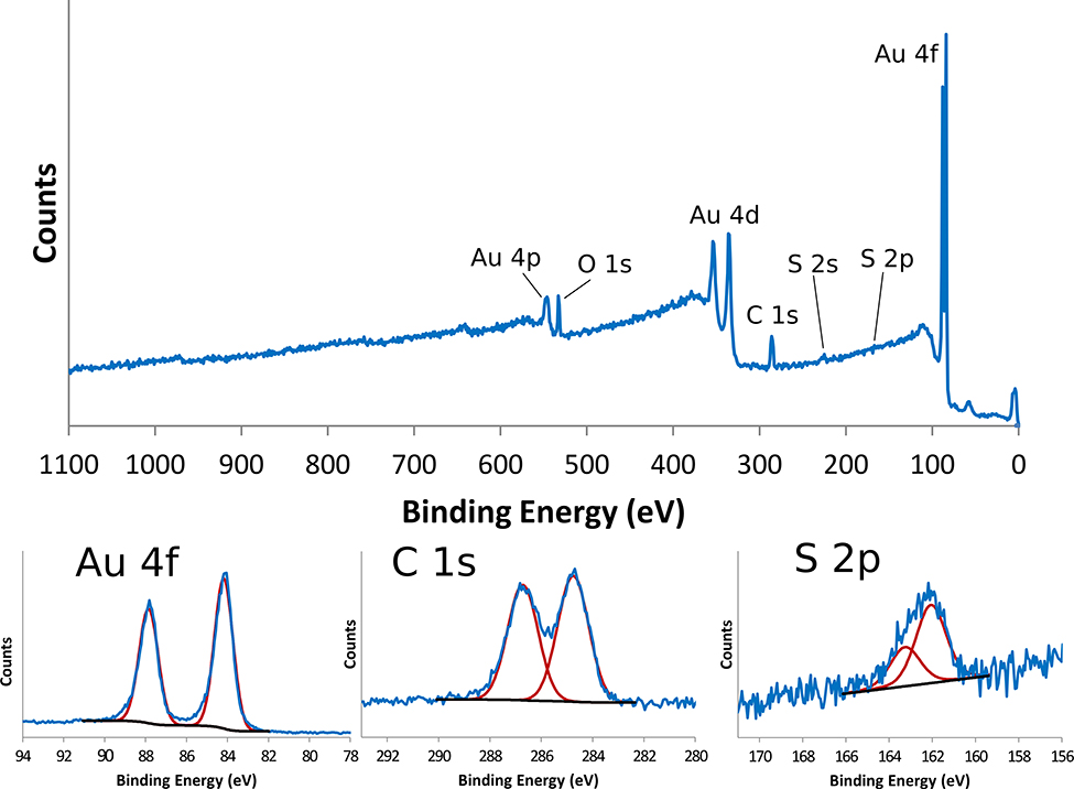
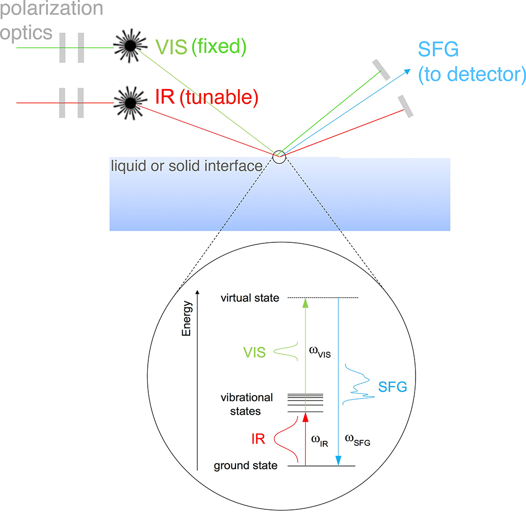
Surfaces represent a unique state of matter that typically have significantly different compositions and structures from the bulk of a material. Since surfaces are the interface between a material and its environment, they play an important role in how a material interacts with its environment. Thus, it is essential to characterize, in as much detail as possible, the surface structure and composition of a material. However, this can be challenging since the surface region typically is only minute portion of the entire material, requiring specialized techniques to selectively probe the surface region. This tutorial will provide a brief review of several techniques used to characterize the surface and interface regions of biological materials. For each technique we provide a description of the key underlying physics and chemistry principles, the information provided, strengths and weaknesses, the types of samples that can be analyzed, and an example application. Given the surface analysis challenges for biological materials, typically there is never just one technique that can provide a complete surface characterization. Thus, a multi-technique approach to biological surface analysis is always required.


Conflict of interest statement

Conflicts of interest

There are no conflicts of interest to declare.

Figures

Figure 1.
Figure 1.
Schematic drawing of a monochromatized XPS instrument with a hemispherical analyzer and multi-channel detector.
Figure 2.
Figure 2.
XPS survey scan (top panel) and XPS high resolution peaks for Au4f (bottom left panel), C1s (bottom middle panel) and S2p (bottom right panel). Red lines show the individual peaks for each peak fit.
Figure 3.
Figure 3.
Schematic drawing of a ToF-SIMS instrument.
Figure 4.
Figure 4.
ToF-SIMS negative ion spectrum from the PEG4thiol SAM.
Figure 5.
Figure 5.
ToF-SIMS negative ion spectra from the PEG4thiol that show the characteristic molecular ion peaks from the PEG4thiol.
Figure 6.
Figure 6.
(a) Schematic of the photoexcitation process that takes place during a NEXAFS experiment. NEXAFS carbon K-edge (b) and XPS C1s (c) spectra acquired from an MHD SAM on gold. NEXAFS carbon K-edge spectra (d) of the PEG4thiol SAM acquired at x-ray incident angles of 90 and 20 degrees from the surface plane.
Figure 7.
Figure 7.
Imaging of an E. gralli head at the carbon K-edge. (a) Photograph of the head of the flower scarab. (b) NEXAFS image of the scarab head. The image is representative of the electron yield across the carbon region 270–370 eV. Each pixel contains a full NEXAFS spectrum. (c) NEXAFS spectra extracted from the image along the line indicated in the image. This figure has been reproduced from reference 31 with permission from Springer Nature, copyright 2019.
Figure 8.
Figure 8.
Schematic of SFG vibrational spectroscopy in reflection mode.
Figure 9.
Figure 9.
SFG spectra taken from PEG4thiol SAMs on Au. Left Panel: SFG C-H region spectra in contact with air (bottom) and deuterated water (top). Prominent peaks are labeled d+ (2846 cm−1), o+ (2891cm−1), and o- (2950 cm−1) which all correspond to symmetric and asymmetric CH2 vibrational modes. Right Panel: The resulting SFG spectra taken as water is exchanged with D2O. Prominent peaks at 3200 and 3400 cm−1 stem from ordered water at the PEG4thiol SAM surface. This figure has been reproduced from reference 37 with permission from American Chemical Society, copyright 2009.
Figure 10.
Figure 10.
Schematic diagrams of wavelength SPR (top) and QCM-D (bottom) biosensing processes.
Figure 11.
Figure 11.
QCM-D sensorgram showing sequential immobilization of Protein G B1 cysteine mutant (V21C), whole IgG and IgG F(ab’)2 fragment onto a maleimide terminated OEG SAM. The green trace represents the frequency change and the gray trace represents the dissipation change.
Figure 12.
Figure 12.
Schematic drawing of a SPM instrument.
Figure 13.
Figure 13.
AFM images collected from a PEG SAM on Au before repeated contact with the AFM tip (panel A) and after 20 distance-force curve measurements (panel B). This figure has been adapted from reference 53 with permission from Elsevier, copyright 2006.

References

    1. Vickerman JC and Gilmore IS, Surface Analysis: The Principal Techniques, John Wiley & Sons, Chichester, UK, 2009.
    1. Castner DG, Biointerphases, 2017, 12, 02C301. - PMC - PubMed
    1. Castner DG, Surface and Interface Analysis, 2018, 50, 981–990. - PMC - PubMed
    1. Castner DG and Ratner BD, Surface Science, 2002, 500, 28–60.
    1. Ratner BD, Weathersby PK, Hoffman AS, Kelly MA and Scharpen LH, Journal of Applied Polymer Science, 1978, 22, 643–664.