. 2020 Jul 30;30(9):4847-4857.
doi: 10.1093/cercor/bhaa077.

Prefrontal Multielectrode Transcranial Direct Current Stimulation Modulates Performance and Neural Activity Serving Visuospatial Processing

Affiliations

Prefrontal Multielectrode Transcranial Direct Current Stimulation Modulates Performance and Neural Activity Serving Visuospatial Processing

Yasra Arif et al. Cereb Cortex. .

Abstract

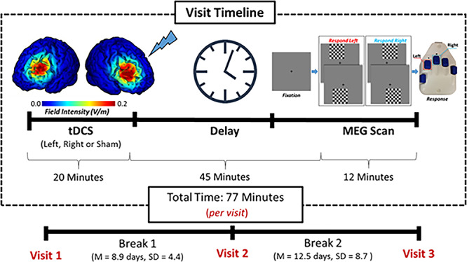
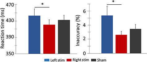
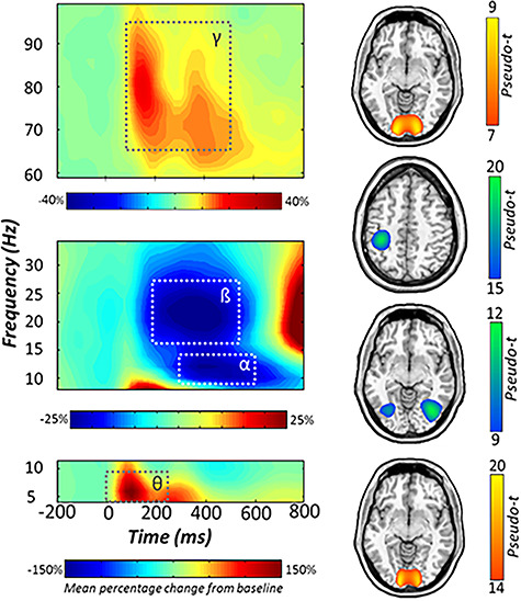
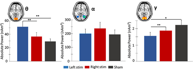
The dorsolateral prefrontal cortex (DLPFC) is known to play a critical role in visuospatial attention and processing, but the relative contribution of the left versus right DLPFC remains poorly understood. We applied multielectrode transcranial direct-current stimulation (ME-tDCS) to the left and right DLPFC to investigate its net impact on behavioral performance and population-level neural activity. The primary hypothesis was that significant laterality effects would be observed in regard to behavior and neural oscillations. Twenty-five healthy adults underwent three visits (left, right, and sham ME-tDCS). Following stimulation, participants completed a visuospatial processing task during magnetoencephalography (MEG). Statistically significant oscillatory events were imaged, and time series were then extracted from the peak voxels of each response. Behavioral findings indicated differences in reaction time and accuracy, with left DLPFC stimulation being associated with slower responses and decreased accuracy compared to right stimulation. Left DLPFC stimulation was also associated with increases in spontaneous theta and decreases in gamma within occipital cortices relative to both right and sham stimulation, while connectivity among DLPFC and visual cortices was generally increased contralateral to stimulation. These data suggest spectrally specific modulation of spontaneous cortical activity at the network-level by ME-tDCS, with distinct outcomes based on the laterality of stimulation.

Keywords: laterality effects; magnetoencephalography; neural oscillations; spontaneous activity.


Figures

Figure 1
Figure 1
Experimental paradigm. Participants received 20 min of anodal and sham ME-tDCS over the left and right DLPFC. Stimulation montages were pseudorandomized across three visits, each separated by at least 1 week. Current distribution modeling revealed focused field intensity values of the left and right DLPFC (left). Following ME-tDCS participants completed a visuospatial paradigm during MEG recording (right). The total visit time from the beginning of stimulation to the end of the MEG task was approximately 77 min, which places the MEG experiment well within the peak sensitivity period for detecting offline effects of ME-tDCS (see Kuo et al. 2013). ME-tDCS, multielectrode transcranial direct current stimulation; DLPFC, dorsolateral prefrontal cortex; MEG, magnetoencephalography.
Figure 2
Figure 2
Behavioral performance on the visuospatial task. Stimulation montage (i.e., left and right active stimulation conditions and sham) is denoted at the bottom with the mean behavioral metrics displayed on the y-axes, with reaction to the left in ms and accuracy to the right in percentage incorrect. Following left DLPFC stimulation, participants were slower and less accurate on the task compared to performance following ME-tDCS of the right DLPFC. Error bars show the SEM. *P < 0.05. ME-tDCS, multielectrode transcranial direct current stimulation; DLPFC, dorsolateral prefrontal cortex; SEM, standard error of the mean.
Figure 3
Figure 3
Neural responses to the visuospatial task. (Left): Grand-averaged time–frequency spectrograms of MEG sensors exhibiting one or more significant responses, with gamma activity at the top, alpha and beta below, and theta at the bottom. In each spectrogram, time (ms) is denoted on the x-axis and frequency (Hz) is shown on the y-axis. All signal power data are expressed as percent difference from baseline, with color legends shown below each respective spectrogram. Dashed lines indicate the time–frequency windows that were subjected to beamforming. (Right): Grand-averaged beamformer images (pseudo-t) across all participants and ME-tDCS montages for each time–frequency component. Axial slices are as follows: Gamma (z = 1), beta (z = 45), alpha (z = −7), and theta (z = −9), from top to bottom. MEG, magnetoencephalography.
Figure 4
Figure 4
Spontaneous theta, alpha, and gamma activity in occipital cortices during the baseline period. Mean absolute power (nAm2) is represented on the y-axes. Repeated measures 1 × 3 ANOVA were computed on spontaneous power averaged over the baseline period (−400 to 0 ms), with active conditions and sham collapsed across the hemispheres. (Left): Elevated theta power was observed following left versus right active and sham stimulation. (Middle): No significant differences were observed in spontaneous alpha power for the three conditions. (Right): Left active stimulation resulted in weaker spontaneous gamma power compared to right active and sham conditions. Error bars reflect the SEM. *P < 0.05. **P < 0.01. SEM, standard error of the mean.
Figure 5
Figure 5
Differential modulation of strength of fronto-visual connectivity by stimulation montages. The glass brains represent functional connections interrogated. Phase locking value (PLV) is represented on the y-axes. Stronger alpha phase coherence was observed between left DLPFC and bilateral visual cortices versus sham following right stimulation. Left active stimulation resulted in higher phase coherence between the right DLPFC and bilateral visual cortices versus sham in alpha and gamma bands. Error bars reflect the SEM. *P < 0.05. SEM, standard error of the mean.

References

    1. Andrews SC, Hoy KE, Enticott PG, Daskalakis ZJ, Fitzgerald PB. 2011. Improving working memory: the effect of combining cognitive activity and anodal transcranial direct current stimulation to the left dorsolateral prefrontal cortex. Brain Stimul. 4:84–89. - PubMed
    1. Balconi M. 2013. Dorsolateral prefrontal cortex, working memory and episodic memory processes: insight through transcranial magnetic stimulation techniques. Neurosci Bull. 29:381–389. - PMC - PubMed
    1. Barbey AK, Koenigs M, Grafman J. 2013. Dorsolateral prefrontal contributions to human working memory. Cortex. 49:1195–1205. - PMC - PubMed
    1. Bikson M, Grossman P, Thomas C, Zannou AL, Jiang J, Adnan T, Mourdoukoutas AP, Kronberg G, Truong D, Boggio P. 2016. Safety of transcranial direct current stimulation: evidence based update 2016. Brain Stimul. 9:641–661. - PMC - PubMed
    1. Boggio PS, Campanhã C, Valasek CA, Fecteau S, Pascual-Leone A, Fregni F. 2010. Modulation of decision-making in a gambling task in older adults with transcranial direct current stimulation. Eur J Neurosci. 31:593–597. - PubMed

Publication types