. 2020 May;61(5):359-370.
doi: 10.3349/ymj.2020.61.5.359.

LncRNA LINC00313 Knockdown Inhibits Tumorigenesis and Metastasis in Human Osteosarcoma by Upregulating FOSL2 through Sponging miR-342-3p

Affiliations

LncRNA LINC00313 Knockdown Inhibits Tumorigenesis and Metastasis in Human Osteosarcoma by Upregulating FOSL2 through Sponging miR-342-3p

Hongtao Chen et al. Yonsei Med J. 2020 May.

Abstract

Purpose: Osteosarcoma (OS) is the most common primary bone tumor, with high morbidity in infants and adolescents. Long noncoding RNA LINC00313 has been found to modulate papillary thyroid cancer tumorigenesis and to be dysregulate in lung cancer. However, the role of LINC00313 in OS has not yet been addressed.

Materials and methods: We evaluated mRNA and protein expression using real-time quantitative PCR and Western blotting. Cell proliferation was evaluated using MTT; apoptosis and autophagy were assessed with flow cytometry, Western blotting, and/or GFP-LC3 assay. Transwell assay was conducted to measure cell migration and invasion. Potential target sites for LINC00313 and miR-342-3p were predicted with starBase v.2.0 and TargetScan Human, and verified using luciferase reporter assay, RNA immunoprecipitation, and RNA pull-down assay. In vivo, xenogeneic tumors were induced with U2OS and MG-63 cells, separately.

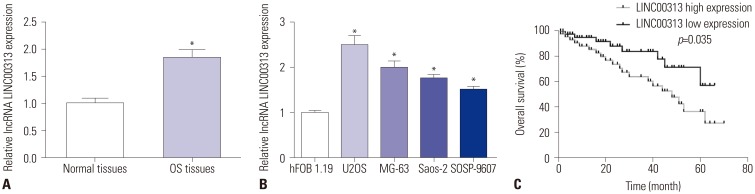
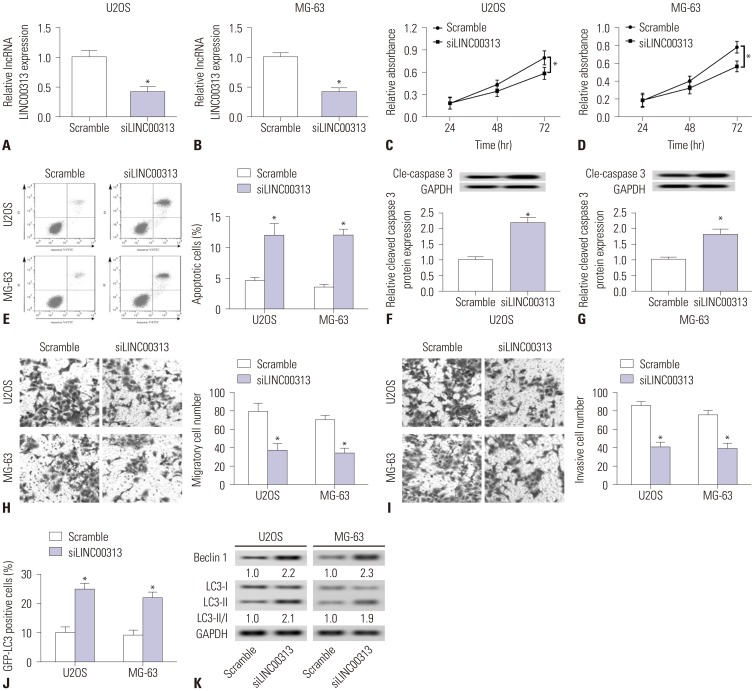
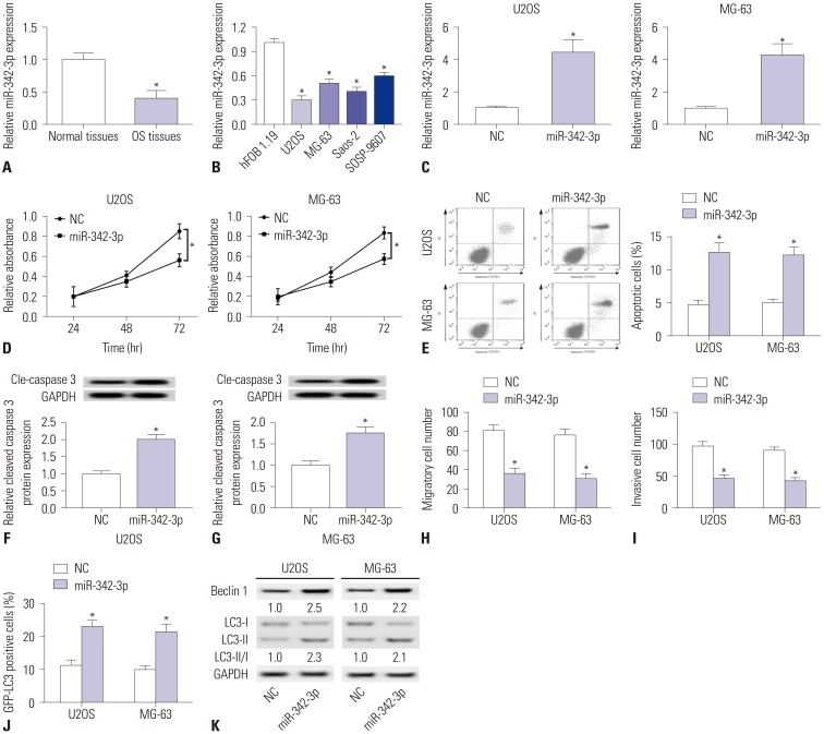
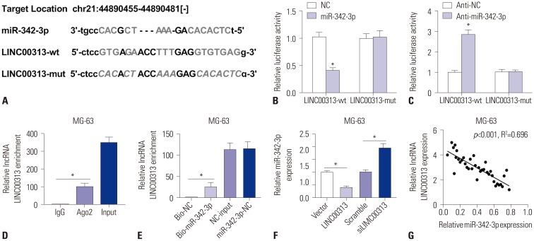
Results: LINC00313 was upregulated and miR-342-3p was downregulated in OS tissues and cells. High expression of LINC00313 was associated with shorter overall survival. FOSL2 downregulation and miR-342-3p overexpression suppressed cell proliferation and migratory and invasive abilities while promoting apoptosis and autophagy, all of which were consistent with the effects of LINC00313 knockdown. miR-342-3p, sponged by LINC00313, inversely modulated FOSL2 by targeting MG-63 cells, and FOSL2 expression was positively controlled by LINC00313. LINC00313 knockdown suppressed tumor growth in vivo.

Conclusion: LINC00313 is upregulated in OS, and LINC00313 knockdown plays a vital anti-tumor role in OS cell progression through a miR-342-3p/FOSL2 axis. Our study suggests that LINC00313 may be a novel, promising biomarker for diagnosis and prognosis of OS.

Keywords: FOSL2; LINC00313; miR-342-3p; osteosarcoma (OS).


Conflict of interest statement

The authors have no potential conflicts of interest to disclose.

Figures

Fig. 1
Fig. 1. Roles of lncRNA LINC00313 in osteosarcoma (OS) tissues and cell lines. (A) Expression of LINC00313 in OS tissues and normal tissues was detected by qPCR. (B) Expression of LINC00313 in OS cells (U2OS, MG-63, Saos-2, and SOSP-9607) and normal osteoblast cells (hFOB 1.19) was detected by qPCR. (C) Survival analysis of differential expression of LINC00313 with Kaplan-Meier curves. All experiments were carried out three times. *p<0.05.
Fig. 2
Fig. 2. Effects of LINC00313 knockdown in osteosarcoma (OS) cell lines. OS cell lines (U2OS and MG-63) were transfected with siLINC00313/Scramble for further study. (A and B) Expression of LINC00313 was detected by qPCR. (C–E) Cell proliferation and apoptosis were measured using MTT staining and flow cytometry. (F and G) Expression of Cle-caspase 3 was detected with Western blotting. (H and I) Cell migration and invasion were assayed with transwell assay (magnification, ×100). (J) GFP-LC3 positive cells were recorded after being transfected with GFP-LC3 plasmid. (K) Expressions of autophagyrelated proteins (Beclin 1, LC3-I, and LC3-II) were detected with Western blotting. GAPDH was the loading control. All experiments were carried out three times. *p<0.05.
Fig. 3
Fig. 3. Roles of miR-342-3p in osteosarcoma (OS) tissues and cell lines. (A) Expression of miR-342-3p in OS tissues and normal tissues was detected by qPCR. (B) Expression of miR-342-3p in OS cells (U2OS, MG-63, Saos-2, and SOSP-9607) and normal osteoblast cells (hFOB 1.19) was detected by qPCR. (C-K) OS cell lines (U2OS and MG-63) were transfected with miR-342-3p mimic (miR-342-3p) and miR-NC mimic (NC) for further study. (C) Expression of miR-342-3p was detected after transfection. (D and E) Cell proliferation and apoptosis were measured by MTT staining and flow cytometry. (F and G) Expression of Cle-caspase 3 was detected with Western blotting. (H and I) Cell migration and invasion were assayed with transwell assay. (J) GFP-LC3 positive cells were recorded after being transfected with GFP-LC3 plasmid. (K) Expressions of Beclin 1, LC3-I, and LC3-II were detected with Western blotting. GAPDH was the loading control. All experiments were carried out three times. *p<0.05.
Fig. 4
Fig. 4. LINC00313 regulates miR-342-3p by targeted binding. (A) Prediction of the potential binding sites between LINC00313 and miR-342-3p on starBase v.2.0. (B and C) Luciferase report assay was performed to determine the luciferase activity of 293T cells transfected between either miR-342-3p/NC or anti-miR-342-3p/anti-NC and LINC00313-wt/LINC00313-mut. (D) RNA immunoprecipitation was conducted to determine LINC00313 expression in MG-63 cell samples. (E) Determination of LINC00313 mRNA in MG-63 cells after transfected with Bio-miR-342-3p/Bio-NC using qPCR. (F) Expression of miR-342-3p in MG-63 cells after being transfected with LINC00313 overexpression plasmid/empty plasmid (LINC00313/Vector), siLINC00313/scrambled siRNA (Scramble), separately. (G) Spearman's correlation analysis between miR-342-3p expression and LINC00313 mRNA levels in 37 OS tissue samples. All experiments were carried out in triplicate. *p<0.05.
Fig. 5
Fig. 5. Effects of restoration of miR-342-3p in osteosarcoma (OS) cell lines. Restoration of miR-342-3p in U2OS and MG-63 cells was induced by co-transfection with siLINC00313 and anti-NC, and defective miR-342-3p restoration was obtained by incubation of siLINC00313 and anti-miR-342-3p. (A) Expression levels of miR-342-3p were detected by qPCR. (B) Cell proliferation was assessed by MTT staining. (C) Cell apoptosis was detected with flow cytometry. (D and E) Activity of caspase 3 was assessed using Western blotting. (F and G) Cell migration and invasion were determined using transwell assay. (H) Positive cells of GFP-LC3 were calculated after transfection. (I) Expressions of Beclin 1, LC3-I, and LC3-II were detected with Western blotting. GAPDH was the loading control. All experiments were carried out in triplicate. *p<0.05.
Fig. 6
Fig. 6. MiR-342-3p modulates osteosarcoma (OS) characteristics by targeting FOSL2. (A) Prediction of potential binding sites between miR-342-3p and FOSL2 on Targetscan Human. (B) Luciferase reporter assay was performed to determine the luciferase activity of 293T cells transfected with miR-342-3p mimics/miR-NC mimic (miR-342-3p/NC) and FOSL2-wt/FOSL2-mut. (C) RNA immunoprecipitation was conducted to determine FOSL2 expressions in MG-63 cell samples. (D and E) Determination of FOSL2 expression both at the mRNA level and protein level in MG-63 cells after transfection with miR-342-3p/NC, anti-miR-342-3p/anti-NC. Restoration of FOSL2 in U2OS and MG-63 cells was induced by co-transfection with miR-342-3p mimic and empty plasmid (Vector), and defective FOSL2 restoration was obtained by incubation of miR-342-3p mimic and FOSL2 overexpression plasmid (FOSL2). (F and G) qPCR detected FOSL2 mRNA expression in OS tissues and cell lines. (H) Cell proliferation was assessed by cell activity with MTT staining. (I) Cell apoptosis was detected with flow cytometry. (J) Activity of caspase 3 was measured using western blotting. (K) Cell migration was determined using Transwell assay. GAPDH was the loading control. (L) Cell invasion was determined using Transwell assay. (M) Positive cells of GFP-LC3 were calculated. (N) Expressions of Beclin 1, LC3-I and LC3-II were detected with western blotting. GAPDH was the loading control. All experiments were carried out in triplicate. *p<0.05.
Fig. 7
Fig. 7. LINC00313 positively regulates FOSL2 by sponging miR-342-3p. Effects of LINC00313 on FOSL2 expression in U2OS and MG-63 cells were illuminated by transfection with siLINC00313/Scramble or co-transfection of siLINC00313 and anti-miR-342-3p/anti-NC. FOSL2 expression was determined using qPCR and Western blotting in U2OS (A and B) and MG-63 (C and D) cells. All experiments were carried out in triplicate. *p<0.05.
Fig. 8
Fig. 8. Effects of LINC00313 knockdown on tumor growth. Lv-ShLINC00313 was infected into U2OS and MG-63 cells, which were implanted in subcutaneous areas of athymic mouse for 25 days. (A and B) Measurement of tumor volume every 5 days, and the weights of tumors were determined on day 25, respectively. (C and D) Detection of LINC00313 expression levels in tumors on day 25. (E and F) Expression levels of miR-342-3p in tumors were measured on day 25. (G and H) Expression levels of FOSL2 protein in tumors were measured on day 25. All experiments were carried out in triplicate. *p<0.05.

References

    1. Yang J, Zhang W. New molecular insights into osteosarcoma targeted therapy. Curr Opin Oncol. 2013;25:398–406. - PubMed
    1. Slade AD, Warneke CL, Hughes DP, Lally PA, Lally KP, Hayes-Jordan AA, et al. Effect of concurrent metastatic disease on survival in children and adolescents undergoing lung resection for metastatic osteosarcoma. J Pediatr Surg. 2015;50:157–160. - PubMed
    1. Yu FX, Hu WJ, He B, Zheng YH, Zhang QY, Chen L. Bone marrow mesenchymal stem cells promote osteosarcoma cell proliferation and invasion. World J Surg Oncol. 2015;13:52. - PMC - PubMed
    1. Fontanella R, Pelagalli A, Nardelli A, D’Alterio C, Ieranò C, Cerchia L, et al. A novel antagonist of CXCR4 prevents bone marrow-derived mesenchymal stem cell-mediated osteosarcoma and hepatocellular carcinoma cell migration and invasion. Cancer Lett. 2016;370:100–107. - PubMed
    1. Pietrovito L, Leo A, Gori V, Lulli M, Parri M, Becherucci V, et al. Bone marrow-derived mesenchymal stem cells promote invasiveness and transendothelial migration of osteosarcoma cells via a mesenchymal to amoeboid transition. Mol Oncol. 2018;12:659–676. - PMC - PubMed

MeSH terms