

Reconstruction of the Midface and Palate
- PMID: 32390774
- PMCID: PMC7202912
- DOI: 10.1055/s-0040-1709470
Reconstruction of the Midface and Palate
Abstract
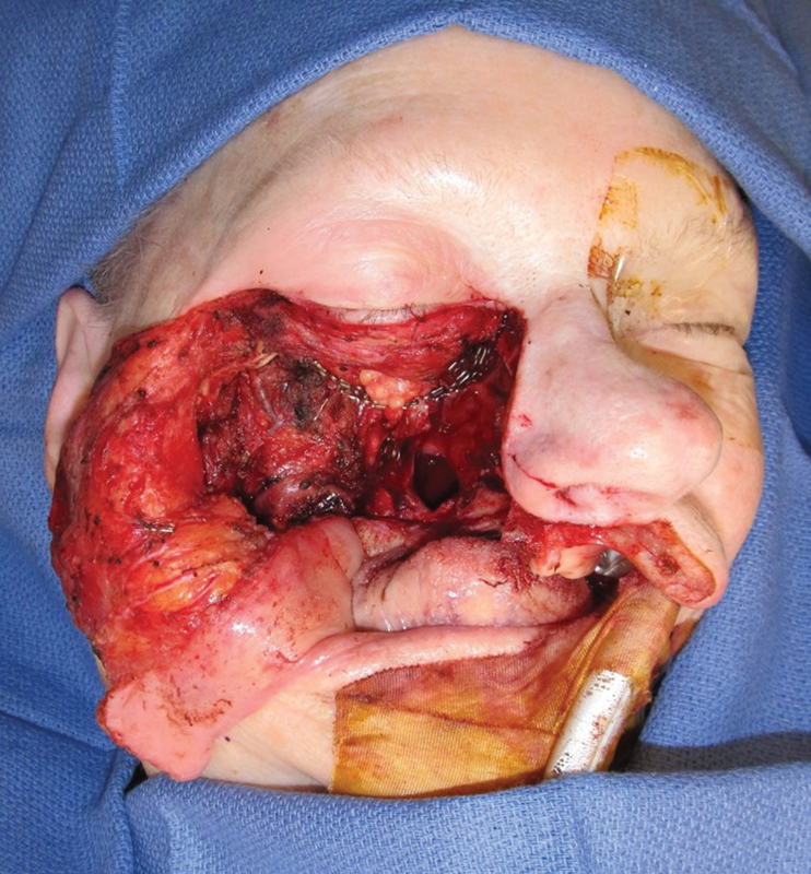
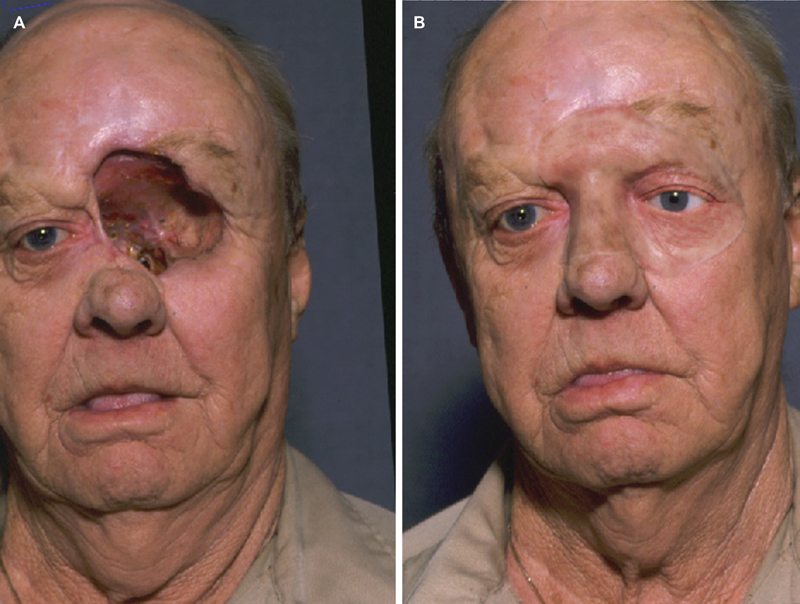
The midface is a complex anatomic structure that is fundamental to many physiologic and homeostatic functions. It may be involved in many pathologic processes that require partial or complete removal. When this happens, reconstruction is mandatory to improve cosmetic outcome with its effect on social interaction as well as to provide an opportunity for complete orodental rehabilitation with restoration of all physiologic functions. This article will review the different reconstructive options available for complex defects of the maxillofacial complex. It will highlight the surgical options available to maximize functional restoration. Finally, it will discuss computer modeling to optimize reconstructive planning.
Keywords: dental rehabilitation; free tissue transfer; maxilla; maxillofacial reconstruction; surgical reconstruction.
© Thieme Medical Publishers.
Conflict of interest statement
Conflict of Interest None declared.
Figures


References
-
- Triana R J, Jr, Uglesic V, Virag M et al. Microvascular free flap reconstructive options in patients with partial and total maxillectomy defects. Arch Facial Plast Surg. 2000;2(02):91–101. - PubMed
-
- Futran N D, Mendez E. Developments in reconstruction of midface and maxilla. Lancet Oncol. 2006;7(03):249–258. - PubMed
-
- Moreno M A, Skoracki R J, Hanna E Y, Hanasono M M. Microvascular free flap reconstruction versus palatal obturation for maxillectomy defects. Head Neck. 2010;32(07):860–868. - PubMed
-
- Futran N D, Haller J R. Considerations for free-flap reconstruction of the hard palate. Arch Otolaryngol Head Neck Surg. 1999;125(06):665–669. - PubMed
-
- Kroll S S, Reece G P, Robb G, Black J. Deep-plane cervicofacial rotation-advancement flap for reconstruction of large cheek defects. Plast Reconstr Surg. 1994;94(01):88–93. - PubMed
