

Modeling Parkinson's Disease With the Alpha-Synuclein Protein
- PMID: 32390826
- PMCID: PMC7191035
- DOI: 10.3389/fphar.2020.00356
Modeling Parkinson's Disease With the Alpha-Synuclein Protein
Abstract
Alpha-synuclein (α-Syn) is a key protein involved in Parkinson's disease (PD) pathology. PD is characterized by the loss of dopaminergic neuronal cells in the substantia nigra pars compacta and the abnormal accumulation and aggregation of α-Syn in the form of Lewy bodies and Lewy neurites. More precisely, the aggregation of α-Syn is associated with the dysfunctionality and degeneration of neurons in PD. Moreover, mutations in the SNCA gene, which encodes α-Syn, cause familial forms of PD and are the basis of sporadic PD risk. Given the role of the α-Syn protein in the pathology of PD, animal models that reflect the dopaminergic neuronal loss and the widespread and progressive formation of α-Syn aggregates in different areas of the brain constitute a valuable tool. Indeed, animal models of PD are important for understanding the molecular mechanisms of the disease and might contribute to the development and validation of new therapies. In the absence of animal models that faithfully reproduce human PD, in recent years, numerous animal models of PD based on α-Syn have been generated. In this review, we summarize the main features of the α-Syn pre-formed fibrils (PFFs) model and recombinant adeno-associated virus vector (rAAV) mediated α-Syn overexpression models, providing a detailed comparative analysis of both models. Here, we discuss how each model has contributed to our understanding of PD pathology and the advantages and weakness of each of them.
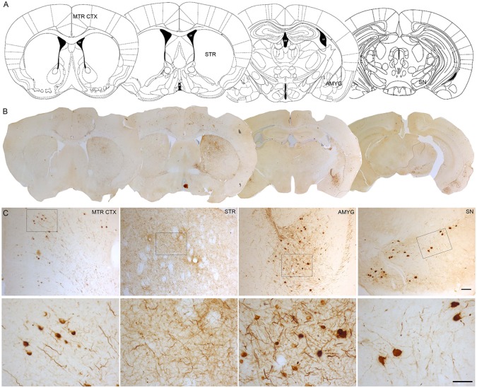
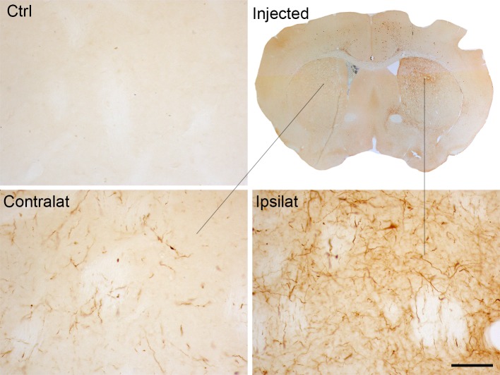
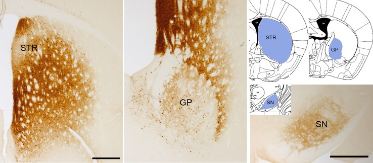
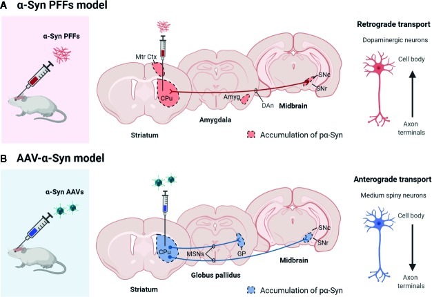
Significance: Here, we show that injection of α-Syn PFFs and overexpression of α-Syn mediated by rAAV lead to a different pattern of PD pathology in rodents. First, α-Syn PFFs models trigger the Lewy body-like inclusions formation in brain regions directly interconnected with the injection site, suggesting that there is an inter-neuronal transmission of the α-Syn pathology. In contrast, rAAV-mediated α-Syn overexpression in the brain limits the α-Syn aggregates within the transduced neurons. Second, phosphorylated α-Syn inclusions obtained with rAAV are predominantly nuclear with a punctate appearance that becomes diffuse along the neuronal fibers, whereas α-Syn PFFs models lead to the formation of cytoplasmic aggregates of phosphorylated α-Syn reminiscent of Lewy bodies and Lewy neurites.
Keywords: Braak hypothesis; Lewy body pathology; prion-like propagation; α-Syn AVV-viral particles; α-Syn aggregation; α-Syn pre-formed fibrils.
Copyright © 2020 Gómez-Benito, Granado, García-Sanz, Michel, Dumoulin and Moratalla.
Figures
References
Publication types
LinkOut - more resources
Full Text Sources
Other Literature Sources
Miscellaneous