. 2020 Apr 23:11:507.
doi: 10.3389/fphar.2020.00507. eCollection 2020.

Anti-Melanogenic Effect of Dendropanax morbiferus and Its Active Components via Protein Kinase A/Cyclic Adenosine Monophosphate-Responsive Binding Protein- and p38 Mitogen-Activated Protein Kinase-Mediated Microphthalmia-Associated Transcription Factor Downregulation

Affiliations

Anti-Melanogenic Effect of Dendropanax morbiferus and Its Active Components via Protein Kinase A/Cyclic Adenosine Monophosphate-Responsive Binding Protein- and p38 Mitogen-Activated Protein Kinase-Mediated Microphthalmia-Associated Transcription Factor Downregulation

Jung Up Park et al. Front Pharmacol. .

Abstract

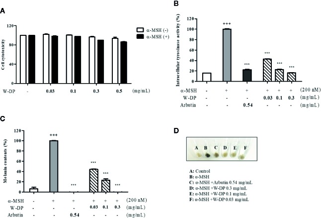
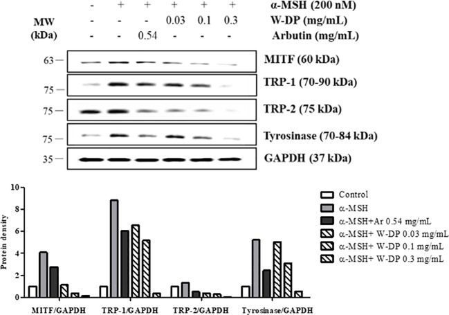
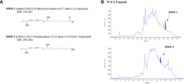
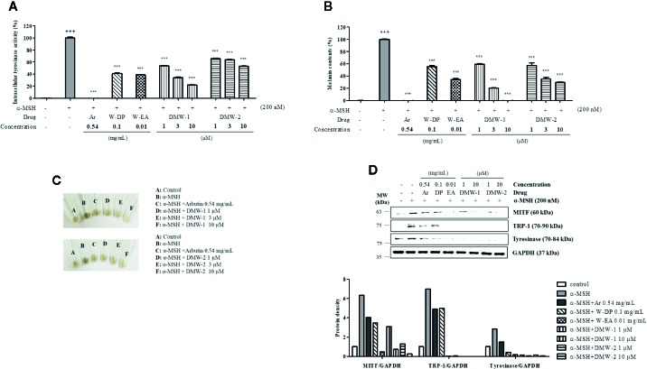
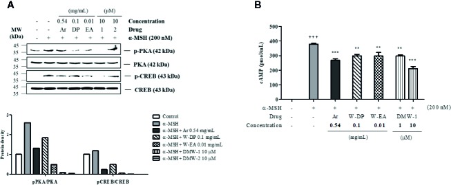
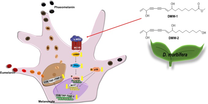
Dendropanax morbiferus H. Lév has been reported to have some pharmacologic activities and also interested in functional cosmetics. We found that the water extract of D. morbiferus leaves significantly inhibited tyrosinase activity and melanin formation in α-melanocyte stimulating hormone (MSH)-induced B16-F10 cells. D. morbiferus reduced melanogenesis-related protein levels, such as microphthalmia-associated transcription factor (MITF), TRP-1, and TRP-2, without any cytotoxicity. Two active ingredients of D. morbiferus, (10E)-9,16-dihydroxyoctadeca-10,17-dien-12,14-diynoate (DMW-1) and (10E)-(-)-10,17-octadecadiene-12,14-diyne-1,9,16-triol (DMW-2) were identified by testing the anti-melanogenic effects and then by liquid chromatography-tandem mass spectrometry (LC/MS/MS) analysis. DMW-1 and DMW-2 significantly inhibited melanogenesis by the suppression of protein kinase A (PKA)/cyclic AMP (cAMP)-responsive binding protein (CREB) and p38 MAPK phosphorylation. DMW-1 showed a better inhibitory effect than DMW-2 in α-MSH-induced B16-F10 cells. D. morbiferus and its active component DMW-1 inhibited melanogenesis through the downregulation of cAMP, p-PKA/CREB, p-p38, MITF, TRP-1, TRP-2, and tyrosinase. These results indicate that D. morbiferus and DMW-1 may be useful ingredients for cosmetics and therapeutic agents for skin hyperpigmentation disorders.

Keywords: (10E)-9,16-dihydroxyoctadeca-10,17-dien-12,14-diynoate; Dendropanax morbiferus; PKA/CREB; anti-melanogenesis; microphthalmia−associated transcription factor; p-p38/p38; tyrosinase.


Figures

Figure 1
Figure 1
Effect of W-DP on melanogenesis in α-MSH-induced B16-F10 cells. B16-F10 cells pretreated with different concentrations of W-DP (0.03, 0.1, 0.3, 0.5 mg/ml) were incubated with or without α-MSH (200 nM) for 72 h. (A) Cell viability was tested by MTS. (B) The cells were then lysed and quantified by using Bradford reagent. Equal amounts of the lysates were treated with L-DOPA (2 mg/ml) in 96-well plate at 37°C for 2 h. The production of DOPAchrome was measured using an ELISA microplate reader at an absorbance of 490 nm. (C) The cells were dissolved in 1 N NaOH with 10% DMSO for 1 h at 60°C. Total melanin lysates were transferred to a 96-well plate and the optical absorbance was measured at 405 nm using an ELISA microplate reader. (D) Colors of the pellets indicated the change of melanogenesis from α-MSH-induced B16-F10 cells. Data are expressed as the mean ± SEM of three independent experiments. *** P < 0.001 versus α-MSH stimulation. +++ P < 0.001 versus the untreated control.
Figure 2
Figure 2
Effect of Dendropanax morbiferus water extract (W-DP) on the expression of melanogenesis-related proteins in α-MSH-induced B16-F10 cells. B16-F10 cells were pretreated with various concentrations of W-DP (0.03, 0.1, 0.3 mg/ml) or arbutin (0.54 mg/ml) for 2 h and then stimulated with α-MSH (200 nM) for 24 h. The protein levels of MITF, tyrosinase, TRP-1, and TRP-2 were detected by Western blot analysis. Arbutin was used as a positive control, and GAPDH was used as the loading control. MITF, tyrosinase, TRP-1, and TRP-2 were analyzed and quantified using Azure Spot software.
Figure 3
Figure 3
Chemical structures and chromatograms of DMW-1 and 2. (A) Chemical structures of DMW-1 and 2 and (B) total ion chromatogram of DMW-1 and 2 in W-EA at 5 mg/ml. W-EA, ethyl acetate fraction.
Figure 4
Figure 4
Effects of DMW-1 and 2 on melanogenesis in α-MSH-induced B16-F10 cells. B16-F10 cells (1 x 10⁵ cells/well) were incubated into six-well plate at 37°C overnight. B16-F10 cells were pretreated with W-DP (0.1 mg/ml), W-EA (0.01 mg/ml), DMW-1 (1, 3, 10 µM), DMW-2 (1, 3, 10 µM), or arbutin (0.54 mg/ml) for 2 h prior to stimulation with α-MSH (200 nM) for 72 h. (A) Equal amounts of the lysates were treated with L-DOPA (2 mg/ml) at 37°C for 2 h. The production of DOPAchrome was measured at an absorbance of 490 nm. (B) The cells were dissolved in 1 N NaOH with 10% DMSO for 1 h at 60°C. Total melanin lysates were measured at an absorbance of 405 nm. (C) Colors of the pellets indicated the change of melanogenesis from α-MSH-induced B16-F10 cells. (D) The expression levels of MITF, tyrosinase, and TRP-1 were determined by Western blot analysis, and the protein bands were quantified using Azure Spot software. GAPDH was used as the loading control. Data are expressed as the mean ± SEM of three independent experiments. ***P < 0.001 versus α-MSH stimulation. +++ P < 0.001 versus the untreated control. Ar, arbutin; W-DP, D. morbiferus water extract; W-EA, ethyl acetate fraction.
Figure 5
Figure 5
Effects of DMW-1 and 2 on the phosphorylation levels of PKA and CREB in α-MSH-induced B16-F10 cells. The cells were pretreated with W-DP (0.1 mg/ml), W-EA (0.01 mg/ml), DMW-1 (10 µM), DMW-2 (10 µM), or arbutin (0.54 mg/ml) for 2 h before stimulation with α-MSH (200 nM) for 24 h. (A) The p-PKA and p-CREB protein levels were determined by Western blot analysis, and normalized to total PKA and CREB. The phosphorylation levels of PKA and CREB were analyzed and quantified using Azure Spot software. (B) The lysates were collected and cAMP levels were tested by ELISA. Data are expressed as the mean ± SEM of three independent experiments. **P < 0.01, and ***P < 0.001 versus α-MSH stimulation. +++ P < 0.001 versus the untreated control. Ar, arbutin; W-DP, D. morbiferus water extract; W-EA, ethyl acetate fraction.
Figure 6
Figure 6
Effect of DMW-1 and 2 on p38 phosphorylation in α-MSH-stimulated B16-F10 cells. B16-F10 cells were pretreated with W-DP (0.1 mg/ml), W-EA (0.01 mg/ml), DMW-1 (10 µM), DMW-2 (10 µM), or arbutin (0.54 mg/ml) for 2 h before stimulation with α-MSH (200 nM) for 24 h. Phosphorylation levels of ERK, JNK, and p38 were determined by Western blot analysis. Equal levels of proteins were confirmed using total ERK, JNK, and p38 antibodies. The protein bands were analyzed and quantified using Azure Spot software. Ar, arbutin; W-DP, D. morbiferus water extract; W-EA, ethyl acetate fraction.
Figure 7
Figure 7
Underlying potential mechanism of anti-melanogenesis of D. morbiferus. The anti-melanogenesis effect of D. morbiferus might be associated with downregulation of MITF. D. morbiferus and its active component DMW-1 significantly inhibited melanin formation by inhibition of melanogenesis-related proteins such as tyrosinase, TRP-1, and TRP-2. D. morbiferus and DMW-1 show multifunctional inhibitory activities on melanogenesis pathways via PKA/CREB- and p38 MAPK-mediated MITF degradation.

Similar articles

Cited by

References

    1. Birhanu B. T., Kim J. Y., Hossain M. A., Choi J. W., Lee S. P., Park S. C. (2018). An in vivo immunomodulatory and anti-inflammatory study of fermented Dendropanax morbifera Leveille leaf extract. BMC Complement Altern. Med. 18, 1-8. 10.1186/s12906-018-2282-x - DOI - PMC - PubMed
    1. Brenner M., Hearing V. J. (2008). The protective role of melanin against UV damage in human skin. Photochem. Photobiol. 84 (3), 539–549. 10.1111/j.1751-1097.2007.00226.x - DOI - PMC - PubMed
    1. Chan C. F., Huang C. C., Lee M. Y., Lin Y. S. (2014). Fermented broth in tyrosinase- and melanogenesis inhibition. Molecules 19 (9), 13122–13135. 10.3390/molecules190913122 - DOI - PMC - PubMed
    1. Cho S. S., Song S. H., Choi C. Y., Park K. M., Shim J. H., Park D. H. (2018). Optimization of the extraction conditions and biological evaluation of Dendropanax morbifera H. Lev as an anti-hyperuricemic source. Molecules 23 (12), 3313–3320. 10.3390/molecules23123313 - DOI - PMC - PubMed
    1. Choo G. S., Lim D. P., Kim S. M., Yoo E. S., Kim S. H., Kim C. H., et al. (2019). Anti−inflammatory effects of Dendropanax morbifera in lipopolysaccharide−stimulated RAW264.7 macrophages and in an animal model of atopic dermatitis. Mol. Med. Rep. 19, 2087–2096. 10.3892/mmr.2019.9887 - DOI - PMC - PubMed