Review
. 2020 Apr 22:11:161.
doi: 10.3389/fendo.2020.00161. eCollection 2020.

General Adaptation in Critical Illness: Glucocorticoid Receptor-alpha Master Regulator of Homeostatic Corrections

Affiliations
Review

General Adaptation in Critical Illness: Glucocorticoid Receptor-alpha Master Regulator of Homeostatic Corrections

Gianfranco Umberto Meduri et al. Front Endocrinol (Lausanne). .

Abstract

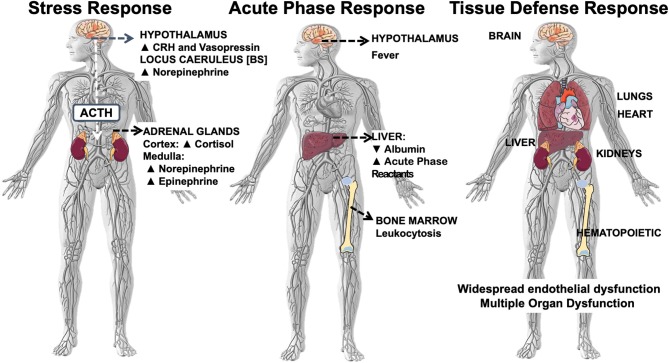
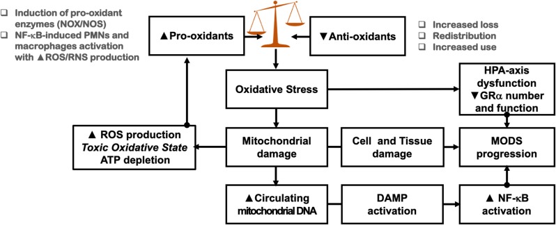
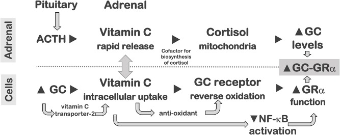
In critical illness, homeostatic corrections representing the culmination of hundreds of millions of years of evolution, are modulated by the activated glucocorticoid receptor alpha (GRα) and are associated with an enormous bioenergetic and metabolic cost. Appreciation of how homeostatic corrections work and how they evolved provides a conceptual framework to understand the complex pathobiology of critical illness. Emerging literature place the activated GRα at the center of all phases of disease development and resolution, including activation and re-enforcement of innate immunity, downregulation of pro-inflammatory transcription factors, and restoration of anatomy and function. By the time critically ill patients necessitate vital organ support for survival, they have reached near exhaustion or exhaustion of neuroendocrine homeostatic compensation, cell bio-energetic and adaptation functions, and reserves of vital micronutrients. We review how critical illness-related corticosteroid insufficiency, mitochondrial dysfunction/damage, and hypovitaminosis collectively interact to accelerate an anti-homeostatic active process of natural selection. Importantly, the allostatic overload imposed by these homeostatic corrections impacts negatively on both acute and long-term morbidity and mortality. Since the bioenergetic and metabolic reserves to support homeostatic corrections are time-limited, early interventions should be directed at increasing GRα and mitochondria number and function. Present understanding of the activated GC-GRα's role in immunomodulation and disease resolution should be taken into account when re-evaluating how to administer glucocorticoid treatment and co-interventions to improve cellular responsiveness. The activated GRα interdependence with functional mitochondria and three vitamin reserves (B1, C, and D) provides a rationale for co-interventions that include prolonged glucocorticoid treatment in association with rapid correction of hypovitaminosis.

Keywords: critical illness; glucocorticoid receptor-alpha; hypovitaminosis; mitochondria; nuclear factor-κB.


Figures

Figure 1
Figure 1
Systemic inflammation-associated multi-system responses: stress, acute phase, tissue defense. Systemic inflammation-associated multi-system response that includes at least three major domains: (i) the stress system composed by the hypothalamic-pituitary-adrenal (HPA) axis and the locus caeruleus-norepinephrine/sympathetic nervous system; (ii) the acute-phase reaction (APR), and (iii) the tissue defense response of the target organs.
Figure 2
Figure 2
Genomic, non-genomic, and mitochondrial glucocorticoid signaling pathways. Glucocorticoid receptor mechanisms of action. The classic actions of GRα are shown in the middle of the panel. The dormant but ligand-friendly receptor, located in the cytoplasm, is bound to co-chaperon molecules, such as heat-shock proteins and the immunophilin FKBP. Upon binding to the ligand, the activated receptor translocates into the nucleus, where it interacts with GREs and/or other transcription factors, such as NF-κB and AP1, to regulate the activity of glucocorticoid-responsive genes, which represent ~20% of the human genome. In addition, cell membrane-associated GRα may be activated by the MAPK and PI3K kinases, as shown in the left of the panel. In addition, by as yet unknown mechanism, the GRα translocates into the mitochondria, where it interacts with the mitochondrial DNA GREs, regulating the activity of mitochondrial genes. See text for additional details cPLA2α, cytosolic phospholipase A2 alpha; eNOS, endothelial nitric oxide synthetase; FKBP, immunophilins; GR, glucocorticoid receptor; HSP, heat shock proteins; MAPK, mitogen-activated protein kinases; NO, nitric oxide; PI3 K, phosphatidylinositol 3-kinase; TF, transcription factor.
Figure 3
Figure 3
Glucocorticoid receptorα as cellular rheostat of homeostatic corrections. The glucocorticoid receptorα (GRα) acts as a cellular rheostat to ensure that a proper response is elicited by the neuroendocrine immune system throughout the three phases of homeostatic corrections: the pro-inflammatory (ready- reinforce), the anti-inflammatory (repress), and the resolution (resolve- restore) phase. Modified with permission from Busillo and Cidlowski (92). TLR2, toll-like receptor 2; purinergic receptor P2Y2R; NLRP3, NOD-like receptor pyrin containing 3; APR, acute phase response; TF, transcription factor; NF-κB, nuclear factor-κB; AP-1, activator protein 1; AnxA1, annexinA1; ALXR, AnxA1 receptor; GILZ, glucocorticoid-induced leucine zipper; TGFβ, transforming growth factor β.
Figure 4
Figure 4
Correlation between mean levels of nuclear NF-κB and nuclear GC-GRα during the natural progression of ARDS, and in response to prolonged glucocorticoid treatment. Mean intracellular changes of nuclear GC-activated GRα and NF-κB observed by exposing PBLs of a healthy volunteer to plasma samples collected longitudinally (days 1, 3, 5, and 8) and after randomization to methylprednisolone treatment [randomization day (R) and post-randomization days 3, 5, 7, and 10]. The mean values of nuclear NF-κB are plotted against the mean nuclear GC-GRα levels. Improvers had a pre-defined improvement in lung injury score (139) and/or gas exchange component by day 7. The left panel shows ARDS patients with adaptive and maladaptive responses. In improvers, an inverse relation was observed between these two transcription factors, with the longitudinal direction of the interaction shifting to the left (decreased NF-κB) and upward (increased GC-GRα). In contrast, in non-improvers NF-κB increased over time while GC-GRα had no significant changes. We define the first interaction as GC-GRα-driven, and the second interaction as NF-κB-driven (33). The right panel shows non-improvers-survivors randomized after day 8 of ARDS to methylprednisolone (n = 11) vs. placebo (n = 6). After natural logarithmic transformation and adjustment for repeated measurements, partial correlations among responses to plasma from the methylprednisolone group were −0.92 (p < 0.0001) both for nuclear NF-κB and nuclear GRα. For responses to plasma from the placebo group, no significant relation was found between nuclear NF-κB and nuclear GRα (r = 0.11; p = 0.70) (103). Reproduced with permission from Meduri et al. (11).
Figure 5
Figure 5
Impact of oxidative stress on homeostatic corrections. See sections Mitochondrial Cacostatic Load, Oxidative Stress, and Mitochondrial Damage, Mitochondria and HPA-Axis Cross-Talk, and Oxidative Stress and CIRCI for explanation. PMN, polymorphonuclear cells; MODS, multiple organ dysfunction syndrome.
Figure 6
Figure 6
Impact of vitamin C on activated GRα activity. (Top) Adrenal: The adrenal and pituitary glands have very high concentrations of ascorbic acid (vitamin C). In response to ACTH release from the corticotrophs of the anterior pituitary gland, the adrenal gland rapidly secretes ascorbic acid, an essential cofactor required for adrenal steroidogenesis in mitochondria, contributing to increased glucocorticoids synthesis. (Bottom) Cells: glucocorticoids, in a time- and concentration-dependent manner, increase the expression of mSVCT2, facilitating the uptake of ascorbic acid into the cell. Ascorbic acid reverses oxidation of the GR, restoring cellular glucocorticoid-responsiveness in oxidant conditions. In addition, ascorbic acid inhibits TNFα- and IL-1β-induced activation of NF-κB in a dose-dependent manner by inhibiting phosphorylation and degradation of IκBα. These combined actions result in increased glucocorticoid availability and GC-GRα activation and improved homeostatic corrections.
Figure 7
Figure 7
Regression of concentration of methylprednisolone on steady-state mRNA levels of TNF-α in U937 cells primed with graded concentrations of lipopolysaccharide (LPS). In experimental simulation of severe inflammation, human monocytic cells (U937 cells) were activated with graded concentrations of LPS (100 ng/ml, 1.0 μg/ml, 5.0 μg/ml, or 10.0 μg/ml) for 6 h followed by measurement of the expression of inflammatory cytokines [TNF-α (shown), IL-1β, and IL-6]. Graded concentrations of LPS were followed by progressively higher inflammatory cytokine transcription (for TNF-α: 185, 318, 481, and 566, respectively). These cells were then exposed to graded concentrations of methylprednisolone [(μg/ml): 50, 100, 150, 200, 250] for 6 h followed by repeated measurement of inflammatory cytokine expression (see below). The steady state mRNA levels of TNF-α, IL-1β, or IL-6 in LPS-activated cells were reduced by treatment with methylprednisolone in a concentration-dependent manner. The effective dose of methylprednisolone was 175 mg, a value that appeared to be independent of the priming level of LPS and type of mRNA measured (102). Modified with permission from Meduri et al. (102).

References

    1. Galluzzi L, Yamazaki T, Kroemer G. Linking cellular stress responses to systemic homeostasis. Nat Rev Mol Cell Biol. (2018) 19:731–45. 10.1038/s41580-018-0068-0 - DOI - PubMed
    1. McEwen BS, Wingfield JC. The concept of allostasis in biology and biomedicine. Horm. Behav. (2003) 43:2–15. 10.1016/S0018-506X(02)00024-7 - DOI - PubMed
    1. Nylen ES, Muller B. Endocrine changes in critical illness. J. Intensive Care Med. (2004) 19:67–82. 10.1177/0885066603259551 - DOI - PubMed
    1. Nesse RM, Young EA. Evolutionary origins and functions of the stress response. J Encyclopedia Stress. (2000) 2:79–84.
    1. Del Rey A, Besedovsky HO. Immune-neuro-endocrine reflexes, circuits, and networks: physiologic and evolutionary implications. Front Horm Res. (2017) 48:1–18. 10.1159/000452902 - DOI - PubMed

MeSH terms

Substances