Case Reports
. 2020 Apr 22:11:235.
doi: 10.3389/fendo.2020.00235. eCollection 2020.

Multidisciplinary Diagnosis of Subcutaneous Soft Tissue Metastasis of Follicular Thyroid Carcinoma: A Case Report

Affiliations
Case Reports

Multidisciplinary Diagnosis of Subcutaneous Soft Tissue Metastasis of Follicular Thyroid Carcinoma: A Case Report

Yanfang Wang et al. Front Endocrinol (Lausanne). .

Abstract

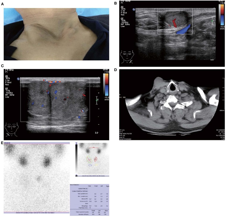
Background: Subcutaneous soft tissue metastasis of follicular thyroid carcinoma (FTC) is rarely diagnosed before surgery for clinicians. Case Report: We present a case of a 67-year-old man with a history of FTC and papillary thyroid microcarcinoma for 5 years. Multiple protruding subcutaneous nodules of the neck were found and removed from the surface of the sternocleidomastoid muscle. Ultrasound, computed tomography and technetium-99 m pertechnetate single-photon emission computed tomography of the neck were performed before the operation, which unfortunately indicated suspicious malignant lesions. Serum Tg was > 300 ng/ml (0.83-68.0 ng/ml), TSH was 36.580 uIU/ml (0.380-4.340 uIU/ml) and AbTg was negative. The pathologic diagnosis was metastatic FTC, invading the surrounding striated muscle, adipose tissue and vessels. Immunohistochemical staining revealed the tumor cells to be positive for thyroglobulin and TTF-1. The specimens of these nodules were further investigated for TERT promoter mutation and the result revealed mutated type (position g 1,295, 228 C>T). Conclusion: Preoperative diagnosis and prognostic prediction of metastatic FTC may be available through a combination of clinical, multimodal imaging and molecular genetic test (viz. multidisciplinary diagnosis). A long-term standardized follow-up is required for patients with a previous diagnosis of FTC.

Keywords: TERT promoter; case report; diagnosis; follicular thyroid carcinoma; metastasis.


Figures

Figure 1
Figure 1
Images of patient and subcutaneous nodules. (A) Multiple subcutaneous soft tissue nodules of metastatic follicular thyroid carcinoma on the anterior neck. (B) Gray-scale ultrasound shows a hypoechoic nodule in the subcutaneous soft tissue of left neck (size were 16 × 11 mm), with wider than taller, well-defined border, irregular margin, perinodular, and intranodular flow and absence of calcification and peripheral halo. (C) Gray-scale ultrasound shows two solid hypoechoic nodules adjacent to each other in the subcutaneous soft tissue of right neck (size of the larger one were 28 × 23 mm), with wider than taller, partially well-defined border, partially regular margin, perinodular and intranodular flow and absence of calcification and peripheral halo. (D) Computed tomography shows several low-density nodules in the subcutaneous of the anterior neck. (E) 99mTcO4-SPECT reveals a shadow of a cold nodule in the right anterior neck region and a shadow of a nodule in the left anterior neck region.
Figure 2
Figure 2
Contrast-enhanced ultrasound and elastosonography appearance of the larger subcutaneous nodules on the bilateral neck. (A,B) Contrast-enhanced ultrasound showed both the larger nodule in the bilateral neck are hypervascular, heterogeneous enhancement, no ring enhancement, and partially clear enhanced boundary. (C,D) Elastosonography displayed both the larger one were mainly composed of blue color (at least 75% of the nodule was covered in blue). (E) 99mTcO4-SPECT whole body scan was performed and showed no abnormal uptake in the whole body.
Figure 3
Figure 3
Follicular thyroid carcinoma. (Hematoxylin and eosin staining of histological slides, × 200).
Figure 4
Figure 4
The TERT promoter testing displayed a mutation type (position g 1,295, 228 C>T). Genomic DNA was isolated from four 5 μm-thick slices of formalin-fixed paraffin-embedded metastatic follicular thyroid carcinoma samples, using a commercial DNA extraction kit (TIANamp FFPE DNA Kit, TIANGEN, Catalog No. DP130608, China), according to the instruction of the manufacturer. The TERT promoter region (2 mutation hot spots—chr5:1,295, 228 C>T and chr5:1,295, 250 C>T) was amplified with a commercial kit (TERT Genetic Mutation Detection Kit, SinoMD, Catalog No. 20090031, China), according to the manufacturer's instructions. The amplified products were sequenced with Applied Biosystems 3,500 Genetic Analyzeror and mutations were recognized on sequencing electropherograms.

Similar articles

References

    1. Sevinc A, Buyukberber S, Sari R, Baysal T, Mizrak B. Follicular thyroid cancer presenting initially with soft tissue metastasis. Jpn J Clin Oncol. (2000) 30:27–9. 10.1093/jjco/hyd007 - DOI - PubMed
    1. Rodrigues G, Ghosh A. Synchronous bony and soft tissue metastases from follicular carcinoma of the thyroid. J Korean Med Sci. (2003) 18:914–6. 10.3346/jkms.2003.18.6.914 - DOI - PMC - PubMed
    1. Farina E, Monari F, Tallini G, Repaci A, Mazzarotto R, Giunchi F, et al. . Unusual thyroid carcinoma metastases: a case series and literature review. Endocr Pathol. (2016) 27:55–64. 10.1007/s12022-015-9410-7 - DOI - PubMed
    1. Madani A, Jozaghi Y, Tabah R, How J, Mitmaker E. Rare metastases of well-differentiated thyroid cancers: a systematic review. Ann Surg Oncol. (2015) 22:460–6. 10.1245/s10434-014-4058-y - DOI - PubMed
    1. Trimboli P, Guglielmi R, Monti S, Misischi I, Graziano F, Nasrollah N, et al. . Ultrasound sensitivity for thyroid malignancy is increased by real-time elastography: a prospective multicenter study. J Clin Endocrinol Metab. (2012) 97:4524–30. 10.1210/jc.2012-2951 - DOI - PubMed

Publication types