. 2020 Apr 23:11:598.
doi: 10.3389/fmicb.2020.00598. eCollection 2020.

Elucidating the Regulon of a Fur - like Protein in Mycobacterium avium subsp. paratuberculosis (MAP)

Affiliations

Elucidating the Regulon of a Fur - like Protein in Mycobacterium avium subsp. paratuberculosis (MAP)

Fernanda Miyagaki Shoyama et al. Front Microbiol. .

Abstract

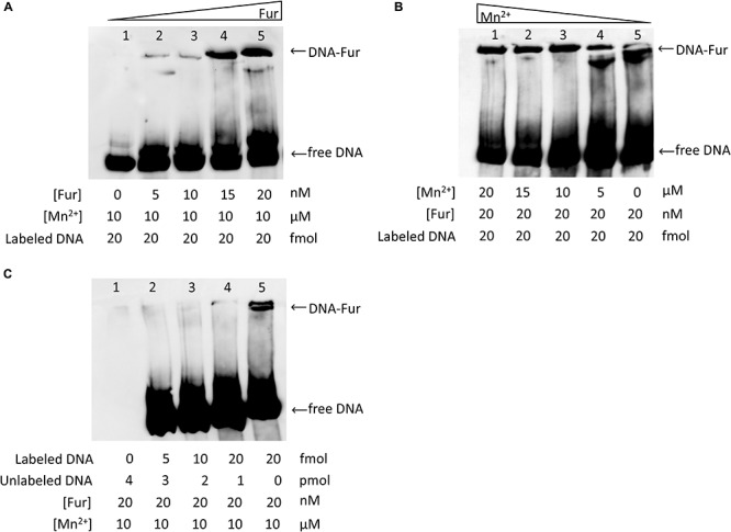
Intracellular iron concentration is tightly regulated to maintain cell viability. Iron plays important roles in electron transport, nucleic acid synthesis, and oxidative stress. A Mycobacterium avium subsp. paratuberculosis (MAP)-specific genomic island carries a putative metal transport operon that includes MAP3773c, which encodes a Fur-like protein. Although well characterized as a global regulator of iron homeostasis in multiple bacteria, the function of Fur (ferric uptake regulator) in MAP is unknown as this organism also carries IdeR (iron dependent regulator), a native iron regulatory protein specific to mycobacteria. Computational analysis using PRODORIC identified 23 different pathways involved in respiration, metabolism, and virulence that were likely regulated by MAP3773c. Thus, chromatin immunoprecipitation followed by high-throughput sequencing (ChIP-seq) was performed to confirm the putative regulon of MAP3773c (Fur-like protein) in MAP. ChIP-Seq revealed enriched binding to 58 regions by Fur under iron-replete and -deplete conditions, located mostly within open reading frames (ORFs). Three ChIP peaks were identified in genes that are directly related to iron regulation: MAP3638c (hemophore-like protein), MAP3736c (Fur box), and MAP3776c (ABC transporter). Fur box consensus sequence was identified, and binding specificity and dependence on Mn2+ availability was confirmed by a chemiluminescent electrophoresis mobility shift assay (EMSA). The results confirmed that MAP3773c is a Fur ortholog that recognizes a 19 bp DNA sequence motif (Fur box) and it is involved in metal homeostasis. This work provides a regulatory network of MAP Fur binding sites during iron-replete and -deplete conditions, highlighting unique properties of Fur regulon in MAP.

Keywords: ChIP-seq; Fur; Mycobacterium avium subsp. paratuberculosis; iron; regulon.


Figures

FIGURE 1
FIGURE 1
In silico analysis of Fur regulon. Using PRODORIC for MAP K-10 genome analysis to detect putative Fur binding and predict pathways regulated by MAP3773c. Solid lines represent pathways directly regulated by MAP3773c. Dashed lines indicated interrelated pathways.
FIGURE 2
FIGURE 2
Identification of MAP Fur protein. (A) Coomassie stain of SDS-PAGE analysis of Fur expression in E. coli system. Lane 1, Protein ladder; Lane 2, BL21(DE3) carrying empty vector pET24b(+); Lane 3, BL21(DE3) carrying MAP3773c on pET24b(+); Lane 4, BL21(DE3) carrying MAP3773c on pET24b(+) with addition of IPTG; Lane 5, Purified recombinant MAP3773c protein. (B) Scaffold analysis of LC-MS/MS data from excised band from lane 5 showing peptide hits (yellow highlights) to 35% of complete MAP Fur sequence. (C) Western blot showing immunoprecipitation of Fur protein by anti-Fur antibody from MAP K-10 cultured under iron-replete and deplete condition. Lane 1, Protein ladder; Lane 2, Pull down of MAP K-10 cultured under iron-replete condition using 1 μg of anti-Fur antibody; Lane 3, Pull down of MAP K-10 cultured under iron-replete condition using 2 μg of anti-Fur antibody; Lane 4, Pull down of MAP K-10 cultured under iron-deplete condition using 1 μg of anti-Fur antibody.
FIGURE 3
FIGURE 3
Overview of the mapped sequences within the reference genome MAP K-10 under iron-replete and -deplete conditions generated by CLC genomic workbench. After mapping onto the reference genome, iron-replete and -deplete samples were compared to control (input DNA), and signal-to-noise (S/N) ratio for peak calling was generated. Fur specifically binds various genomic loci under both conditions, but most of the ChIP peaks showed higher binding sites under iron-replete condition. Arrows indicate regions where ChIP peaks are associated with iron regulation.
FIGURE 4
FIGURE 4
Applying FDR ≤ 10– 50, there are three ChIP peaks associated with iron regulation. (A) MAP Fur protein binds to the region of MAP3638c; however, only under iron-deplete condition is binding statically significant with a peak score of 17.4 (MAP3638c). (B) Under iron-replete conditions, there is a strong binding of MAP Fur to the region of MAP3776c represented by a peak score of 32.51.
FIGURE 5
FIGURE 5
MAP Fur box analysis. (A) The most significant motif derived from ChIP-seq binding sequence using MEME. Height of each letter represents the relative frequency of each base at a different position in the consensus sequence. (B,C) A zoom-in of the MAP Fur boxes generated by CLC genomics. (B) Under both iron conditions, there is no binding of MAP Fur to the region of Fur box 3 (MAP3739c). ChIP peak (9.46) outside the ORF has FDR higher than the threshold of FDR ≤ 10– 50 (C) The enriched region of MAP Fur binding onto Fur Box 1 and 2 identified by ChIP-seq. ChIP peak showed higher occupancy under iron-deplete condition in the Fur Box 1 region. S/N denotes the signal-to-noise ratio for peak calling generated by CLC software.
FIGURE 6
FIGURE 6
EMSA analysis of MAP Fur binding to Fur box consensus DNA. Binding activity is represented by band intensity. Twenty femtomoles of MAP DNA including the Fur Box 1 consensus biotin labeled was run in a 5% native polyacrylamide gel with different concentrations of MAP Fur protein and Mn2+. (A) Protein–DNA binding is dose-dependent: titration of purified MAP Fur protein shows an increase of binding activity as more protein is added to the system. (B) Binding activity is more efficient in the presence of Mn2+: No addition of Mn2+ (Lane 5) binding occurs with a lower band intensity when compared to the sample with Mn2+ (Lanes 1–4). (C) Competitive EMSA. Fur protein was incubated with either biotin-labeled DNA probe or unlabeled DNA probe or with both. Biotin-labeled probe was detected using chemiluminescence-based nucleic acid detection kit. Addition of unlabeled DNA affects binding activity, showing binding specificity.
FIGURE 7
FIGURE 7
Model for the regulation of the iron stimulon in M. paratuberculosis. Under low-iron conditions, the iron sensor MAP3737 (yellow teardrop) initiates a signal transduction cascade activating a hypothetical master regulator (MR) of the stimulon (orange rectangle) leading to the transcription of apo-Fur and apo-IdeR. Under low iron, apo-Fur activates transcription of the iron uptake protein or system (blue oval) that is transported to the cell membrane and carries ferric iron bound to carboxymycobactin (cMyco) (blue cloud) into the bacterium. The cMyco + Fe3+ complex possesses FAD-binding activity, allowing interaction with an iron flavin reductase that converts Fe3+ to Fe2+ and disassociates the complex, liberating Fe2+. Apo-IdeR is inactive but, bound to iron (IdeR Fe2), represses transcription of iron import/export protein or system and iron transport is shut down (red X). In addition, bound to ferrous iron, either regulator can exert a positive (pointed blue arrows) or negative regulation (flat-headed arrows) on the transcription of genes in their corresponding regulons. Apo-Fur also exerts a regulatory effect on the Fur regulon. Some genes may be controlled by both Fur and IdeR in opposite ways (broken blue arrows). More speculative effects are depicted by arrows with question marks. Thus, in this model, both Fur and IdeR act in a coordinate fashion to regulate the iron stimulon composed of the Fur and IdeR regulons. Black pointed arrows are used for processes unrelated to transcription such as binding or signal transduction effects.

Similar articles

Cited by

References

    1. Alexander D. C., Turenne C. Y., Behr M. A. (2009). Insertion and deletion events that define the pathogen Mycobacterium avium subsp. paratuberculosis. J. Bacteriol. 191 1018–1025. 10.1128/JB.01340-08 - DOI - PMC - PubMed
    1. Bagg A., Neilands J. B. (1987). Ferric uptake regulation protein acts as a repressor, employing iron (II) as a cofactor to bind the operator of an iron transport operon in Escherichia coli. Biochemistry 26 5471–5477. 10.1021/bi00391a039 - DOI - PubMed
    1. Bannantine J. P., Baechler E., Zhang Q., Li L., Kapur V. (2002). Genome scale comparison of Mycobacterium avium subsp. paratuberculosis Reveals potential diagnostic sequences. J. Clin. Microbiol. 40 1303–1310. 10.1128/JCM.40.4.1303-1310.2002 - DOI - PMC - PubMed
    1. Bannantine J. P., Paustian M. L. (2006). Identification of Diagnostic Proteins in Mycobacterium avium subsp. paratuberculosis by a Whole Genome Analysis Approach. Diagnostic Bacteriology- Protocol – Second Edition. New Jersey: Humana Press. - PubMed
    1. Bereswill S., Greiner S., van Vliet A. H., Waidner B., Fassbinder F., Schiltz E., et al. (2000). Regulation of ferritin-mediated cytoplasmic iron storage by the ferric uptake regulator homolog (Fur) of Helicobacter pylori. J. Bacteriol. 182 5948–5953. 10.1128/jb.182.21.5948-5953.2000 - DOI - PMC - PubMed

LinkOut - more resources