Review
. 2020 Apr 24:11:660.
doi: 10.3389/fimmu.2020.00660. eCollection 2020.

Structural Insights Into How Proteoglycans Determine Chemokine-CXCR1/CXCR2 Interactions: Progress and Challenges

Affiliations
Review

Structural Insights Into How Proteoglycans Determine Chemokine-CXCR1/CXCR2 Interactions: Progress and Challenges

Krishna Rajarathnam et al. Front Immunol. .

Abstract

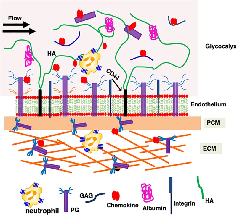
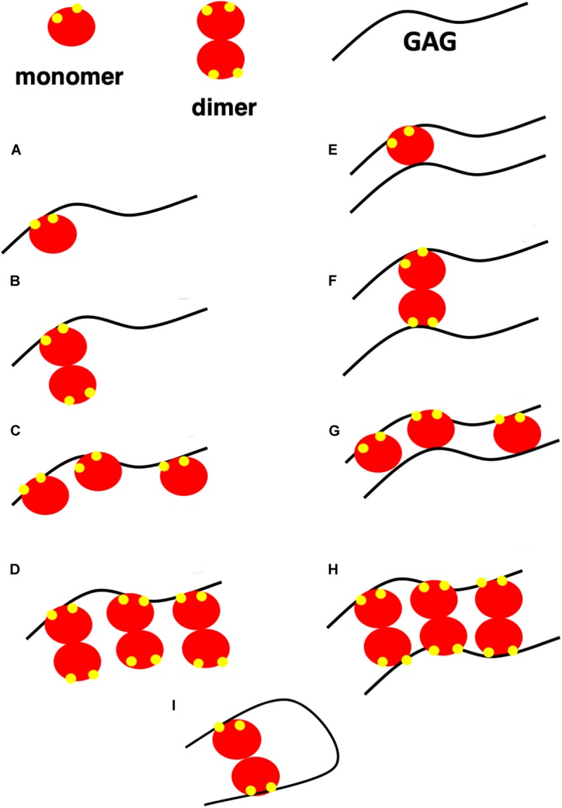
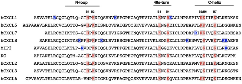
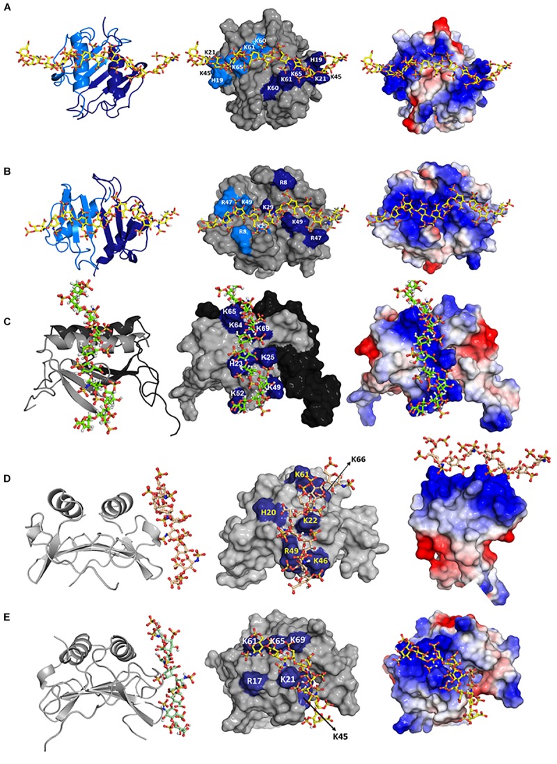
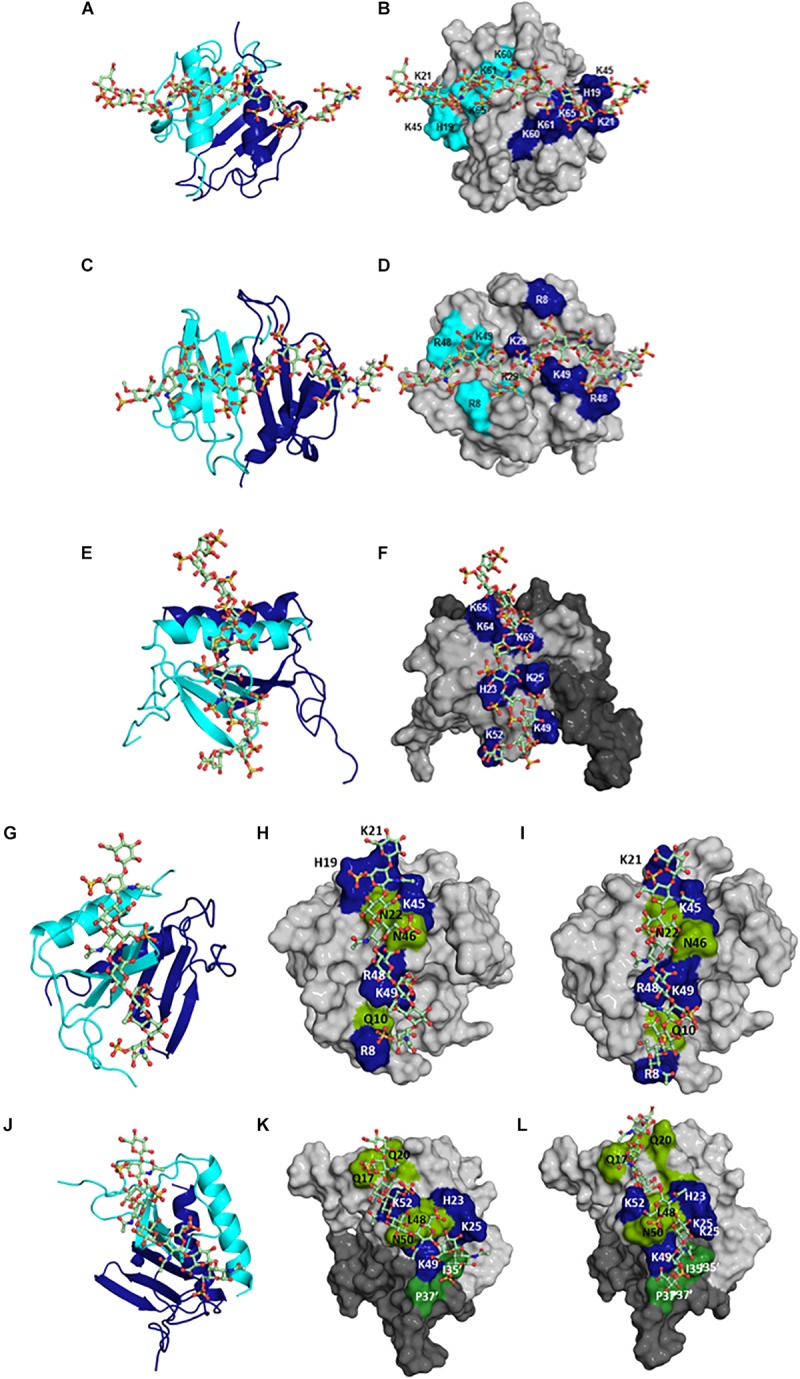
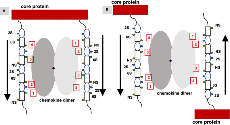
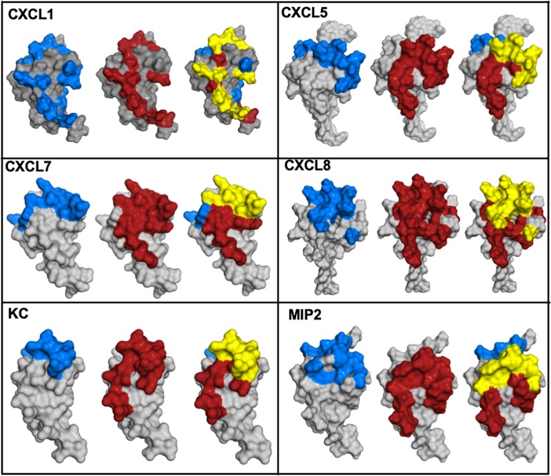
Proteoglycans (PGs), present in diverse environments, such as the cell membrane surface, extracellular milieu, and intracellular granules, are fundamental to life. Sulfated glycosaminoglycans (GAGs) are covalently attached to the core protein of proteoglycans. PGs are complex structures, and are diverse in terms of amino acid sequence, size, shape, and in the nature and number of attached GAG chains, and this diversity is further compounded by the phenomenal diversity in GAG structures. Chemokines play vital roles in human pathophysiology, from combating infection and cancer to leukocyte trafficking, immune surveillance, and neurobiology. Chemokines mediate their function by activating receptors that belong to the GPCR class, and receptor interactions are regulated by how, when, and where chemokines bind GAGs. GAGs fine-tune chemokine function by regulating monomer/dimer levels and chemotactic/haptotactic gradients, which are also coupled to how they are presented to their receptors. Despite their small size and similar structures, chemokines show a range of GAG-binding geometries, affinities, and specificities, indicating that chemokines have evolved to exploit the repertoire of chemical and structural features of GAGs. In this review, we summarize the current status of research on how GAG interactions regulate ELR-chemokine activation of CXCR1 and CXCR2 receptors, and discuss knowledge gaps that must be overcome to establish causal relationships governing the impact of GAG interactions on chemokine function in human health and disease.

Keywords: chemokine; chondroitin sulfate; glycosaminoglycan; heparan sulfate; heparin; nuclear magnetic resonance; proteoglycan; structure.


Figures

FIGURE 1
FIGURE 1
All ELR-chemokines share the same structural fold. Structures of CXCL5 dimer (A) and monomer (B) as a representative of ELR chemokines are shown. The individual monomers in the dimer are shown in dark and light blue for clarity and different GAG-binding regions (N-terminal loop, 40s turn, and C-terminal α-helix) are labeled.
FIGURE 2
FIGURE 2
A schematic showing how proteoglycan interactions regulate chemokine-mediated neutrophil trafficking. Endothelial glycocalyx consists of a number of secreted PGs and various proteins such as albumin that functions as a barrier between blood flow and the endothelium. Critical components of the endothelium and glycocalyx are labeled. This figure is not drawn to scale, and its purpose is to illustrate different anatomical regions that are involved in chemokine-mediated neutrophil recruitment to the insult site.
FIGURE 3
FIGURE 3
A schematic showing possible GAG-bound ELR chemokine structures. In this schematic, a GAG corresponds to heparin or HS, monomer and dimer corresponds to CXCL5 monomer and dimer, and GAG-binding residues are shown in yellow. (A,B) A single chemokine monomer or dimer binding a single GAG occurs at low chemokine concentrations. With increasing concentration, dimer-bound GAG is favored due to higher binding affinity. (C,D) Chemokines bind GAGs like beads on a string at high chemokine concentrations. Of the two, model (D) is favored due to higher binding affinity of the dimer. (E–H) Chemokines bind two GAGs within a PG or between PGs. Of the different models, models E and G are unlikely as a monomer has only one GAG binding site. (I) Horseshoe model of HS binding to a chemokine dimer. HS structure consists of sulfated regions (NS) interspersed with non-sulfated regions (NA). HS is of the form NS-NA-NS in the horseshoe model.
FIGURE 4
FIGURE 4
GAG-binding residues in human and mouse ELR-chemokines. A GAG-binding residue is labeled as conserved if present in five or more of the seven human sequences. Conserved GAG-binding residues are labeled B1 to B7 and are in red. Binding residues unique to a given chemokine are in blue.
FIGURE 5
FIGURE 5
Structural models of heparin bound to (A) CXCL1 α-domain, (B) CXCL1 β-domain, (C) CXCL5, (D) KC/mCXCL1, and (E) MIP2/mCXCL2. Left column: chemokine dimer structures are shown in ribbon presentation and heparin as sticks. Middle column: Heparin binding residues are highlighted on a space-filling model. Right column: Heparin binding residues are shown as the electrostatic surface. In panels (A,B), two monomers of the dimer are shown in light and dark blue (left column) and heparin-binding residues from both monomers are highlighted in light and dark blue (middle column). In panel (C), two monomers of the dimer are shown in gray and black (left and middle columns), and GAG-binding residues are labeled only in the gray monomer. In panels (D,E), both monomers are shown in gray, and only one monomer of the dimer is labeled.
FIGURE 6
FIGURE 6
Structural models of HS bound to (A,B) CXCL1 α-domain, (C,D) CXCL1 β-domain, and (E,F) to CXCL5; of CS bound to (G,H) CXCL1 γ-domain and to (J,K) CXCL5; of DS bound to (I) CXCL1 γ-domain and to (L) CXCL5. In panels (A,C,E,G,J), CXCL1 and CXCL5 dimer structures are shown in ribbon presentation and GAG as sticks. Two monomers of the dimer are shown in blue and cyan. In panels (B,D,F,H,I,K,L), GAG binding residues are highlighted on a space-filling model. In panels (F,K,L), two monomers of the dimer are colored in light and dark gray. In panels (H–K), polar residues are in green. In panels (K,L), residues from the second monomer are distinguished by the ′ symbol. HS binds across the dimer interface in CXCL1 and within the monomer in CXCL5; CS and DS bind within a monomer in CXCL1 and bind both monomers of the dimer in CXCL5.
FIGURE 7
FIGURE 7
Binding of a chemokine dimer to PG GAGs. Monomers of the chemokine dimer are shown in different shades of gray. For illustrative purposes, GAG-binding residues in each monomer of the chemokine dimer are labeled 1 to 4, which run in opposite directions because of two-fold symmetry about the dimer interface (shown by a black dot). Schematic of an heparin octasaccharide with iduronate and glucosamine shown as an oval and rectangle, and N-sulfate (NS), 2-sulfate (2S), and 6-sulfate (6S) shown as spheres in different colors. Helical structure of heparin clusters the 3 sulfate groups on opposite faces of the helical axis (44). (A) Binding of a chemokine dimer to two GAG chains within a PG. Direction of the GAG orientation are shown by arrows. The schematic shows GAG interactions of the two monomers cannot be the same, and that binding to both GAG chains is only possible if the binding interactions of the two monomers are different. (B) Binding of a chemokine to two GAG chains from two PGs. Direction of the GAG orientation are shown by arrows. In this case, two monomers of the dimer can encounter GAG chains running in opposite directions and so similar interactions to both monomers are possible. Such binding could occur in PCM and ECM due to some degree of motional freedom of the PGs.
FIGURE 8
FIGURE 8
Chemokine structures showing GAG and receptor binding regions. Receptor-binding domains are in red, GAG-binding domains are in blue, and residues that are common to both are in yellow.
FIGURE 9
FIGURE 9
Presentation of GAG-bound chemokine for receptor interactions. Chemokine is shown as a red sphere, and shared residues for GAG and receptor binding are shown in orange. (A) Chemokine monomer bound to the receptor cannot bind the GAG. (B) Chemokine monomer bound to a GAG chain cannot bind the receptor. (C) A chemokine dimer bound to a single GAG chain. In this case, one monomer can bind the GAG and the second monomer of the dimer is available for receptor interactions.
FIGURE 10
FIGURE 10
A schematic of the endothelial syndecans and glycocalyx. Syndecans span the membrane and exist as dimers. Sdc-1 is larger (33 kDa) compared to Sdc-4 (22 kDa), and the extracellular domains carry HS and CS chains that are shown in blue and orange, respectively. Chemokine dimer bound to HS chain on syndecans (A), and to free HS and syndecan ectodomain in the glycocalyx (B,C) are accessible to the CXCR2 receptor on neutrophils.

References

    1. Iozzo RV, Schaefer L.Proteoglycan form and function: a comprehensive nomenclature of proteoglycans. Matrix Biol. (2015) 42:11–55. 10.1016/j.matbio.2015.02.003 - DOI - PMC - PubMed
    1. Karamanos NK, Piperigkou Z, Theocharis AD, Watanabe H, Franchi M, Baud S, et al. Proteoglycan chemical diversity drives multifunctional cell regulation and therapeutics. Chem Rev. (2018) 118:9152–232. 10.1021/acs.chemrev.8b00354 - DOI - PubMed
    1. Couchman JR.Transmembrane signaling proteoglycans. Annu Rev Cell Dev Biol. (2010) 26:89–114. 10.1146/annurev-cellbio-100109-104126 - DOI - PubMed
    1. Weinbaum S, Tarbell JM, Damiano ER.The structure and function of the endothelial glycocalyx layer. Annu Rev Biomed Eng. (2007) 9:121–67. 10.1146/annurev.bioeng.9.060906.151959 - DOI - PubMed
    1. Schaefer L, Schaefer RM.Proteoglycans: from structural compounds to signaling molecules. Cell Tissue Res. (2010) 339:237–46. 10.1007/s00441-009-0821-y - DOI - PubMed

Publication types

LinkOut - more resources