Clinical Trial
. 2020 Apr 24:11:666.
doi: 10.3389/fimmu.2020.00666. eCollection 2020.

Necrostatin-1 Ameliorates Neutrophilic Inflammation in Asthma by Suppressing MLKL Phosphorylation to Inhibiting NETs Release

Affiliations
Clinical Trial

Necrostatin-1 Ameliorates Neutrophilic Inflammation in Asthma by Suppressing MLKL Phosphorylation to Inhibiting NETs Release

X A Han et al. Front Immunol. .

Abstract

Neutrophilic inflammation occurs during asthma exacerbation, and especially, in patients with steroid-refractory asthma, but the underlying mechanisms are poorly understood. Recently, a significant accumulation of neutrophil extracellular traps (NETs) in the airways of neutrophilic asthma has been documented, suggesting that NETs may play an important role in the pathogenesis. In this study, we firstly demonstrated that NETs could induce human airway epithelial cell damage in vitro. In a mouse asthmatic model of neutrophil-dominated airway inflammation, we found that NETs were markedly increased in bronchoalveolar lavage (BAL), and the formation of NETs exacerbated the airway inflammation. Additionally, a small-molecule drug necrostatin-1 (Nec-1) shown to inhibit NETs formation was found to alleviate the neutrophil-dominated airway inflammation. Nec-1 reduced total protein concentration, myeloperoxidase activity, and the levels of inflammatory cytokines in BAL. Finally, further experiments proved that the inhibition of Nec-1 on NETs formation might be related to its ability to inhibiting mixed lineage kinase domain-like (MLKL) phosphorylation and perforation. Together, these results document that NETs are closely associated with the pathogenesis of neutrophilic asthma and inhibition of the formation of NETs by Nec-1 may be a new therapeutic strategy to ameliorate neutrophil-dominated airway inflammation.

Keywords: MLKL; Necrostatin-1; asthma; neutrophil extracellular traps; neutrophilic inflammation.


Figures

Figure 1
Figure 1
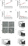
Sputum extracellular DNA (eDNA) levels are significantly correlated with sputum neutrophils in asthmatics and chronic cough patients, and neutrophil extracellular DNA traps (NETs) induced damage of human airway epithelial cells. (A,B) Extracellular DNA in sputum stained with Sytox Green. Sputum eDNA levels were significantly correlated with sputum neutrophils percentage in asthmatics and chronic cough. (C,D) Sputum eDNA levels were not significantly correlated with sputum eosinophil percentage in asthmatics and chronic cough. (E) Morphological changes of 16HBE cells after treatment with 500 ng/ml of NETs for 24 h. 16HBEs were treated with different concentrations of NETs for 24 h, and LDH release (F), cell viability (G), detached cells (%) (H) and supernatant interleukin (IL)-1β (I) were detected. The data are shown as mean ± SD. (*p < 0.05, ***p <0.001, ns, not significant). The results are representative of at least three experiments.
Figure 2
Figure 2
NETs aggravated neutrophil-dominated airway inflammation. (A) Schematic diagram of OVA/CFA sensitized neutrophil-dominated airway inflammation model. (B) Schematic diagram of OVA/CFA plus PMA sensitized neutrophil-dominated airway inflammation model. (C) Pulmonary inflammatory cell populations of OVA/CFA sensitized mice with neutrophil-dominated airway inflammation. (D) The percentages of neutrophils in total BAL cells of OVA/CFA and OVA/CFA plus PMA groups. (E,F) Extracellular DNA in BAL stained with Sytox Green. (G) Representative images of H&E-stained lung tissues of OVA/CFA and OVA/CFA plus PMA groups. Values are expressed as means ± SEM of three independent experiments. (**p < 0.01, ***p < 0.001).
Figure 3
Figure 3
Nec-1 inhibited PMA-induced NETs formation in vitro. (A) Levels of extracellular DNA released by peripheral blood neutrophils, which were cultured with PMA (25 nM) or different concentrations of Nec-1 (50 or 100 μM), or both. (B) Representative immunofluorescence images of Sytox Green-stained neutrophils cultured for 3 h in the absence (ctrl) or presence of PMA (25 nM), Nec-1 (50 μM), or both (200×). (C) Representative immunofluorescence images of neutrophils pretreated with Nec-1 (50 μM) and stimulated with or without PMA (25 nM). Upper panels illustrate DAPI (blue), central panels illustrate MPO (green), lower panels show merged images (400×). (D) Representative immunofluorescence images of neutrophils pretreated without (ctrl) or with Nec-1(50 μM) and stimulated with PMA (25 nM) or both. Upper panels illustrate Hoechst-33342 (blue), central panels illustrate Sytox Green, and lower panels show merged images (200×). The values were shown as mean ± SEM. (**p < 0.01, ****p < 0.0001, ns, not significant). The results are representative of three independent experiments.
Figure 4
Figure 4
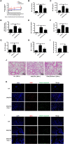
Nec-1 attenuated OVA/CFA sensitized neutrophil-dominated airway inflammation by inhibiting NETs formation. OVA/CFA sensitized mice were treated with Nec-1 (6 mg/kg body weight), and BAL harvested. (A) OVA/CFA sensitized mice were challenged with different doses of methacholine and treated with Nec-1 (6 mg/kg body weight), and BAL and lung tissues were harvested. Airway hyper-responsiveness used to assess the responses of mice to different doses of methacholine. (B) Total number of cells in BAL. (C) MPO activity in lung tissue homogenate. (D) Levels of extracellular DNA in BAL stained with Sytox Green. (E) Total protein concentration in BAL. (F–H) Levels of TNF-α, IFN-γ, and IL-1β in BAL. (I) Fraction of apoptotic neutrophils in the BAL determined by flow cytometry. Quantitative analysis of flow cytometry data (n ≥ 3; ***P < 0.001 vs. control). (J) Representative images of H&E-stained lung tissue of mice from different experimental groups. (J) Representative immunofluorescence images of BAL cells. From left to right, the panels illustrate staining with DAPI, MPO antibody, and cleaved caspase-3 antibody. The right panels show the merge of images (200×). (K,L) Representative immunofluorescence images of BAL cells and lung tissues of control, OVA/CFA, and Nec-1 groups (200×). From left to right, the panels illustrate staining with DAPI, MPO antibody, and cleaved caspase-3 antibody. The right panels show the merge of image. Values are expressed as means ± SEM. (*p < 0.05, **p < 0.01, ***p < 0.001, ns, not significant).
Figure 5
Figure 5
Nec-1 inhibited PMA-induced NETs formation by suppressing phosphorylation and perforation of MLKL in vitro. (A,B) Sytox Green stained neutrophils, stimulated with 25 nM PMA alone, or with different concentrations of Nec-1 (50 or 100 μM) and imaged for 4 h using an automatic live cell imaging analysis system (Videos S1–S4 respectively indicated group Ctrl,PMA (25 nM), PMA+Nec-1 (50 μM) and PMA+ (100 μM) were shown in additional files). (A) showed light field channel (measuring changes in cell morphology and structure under ordinary optics microscope). (B) showed FITC green channel (measuring non-transmembrane nucleic acid dye Sytox Green). The green fibrin-like structure indicated NETs. (C) The mean fluorescence intensity of Sytox Green at different time points analyzed by Image J semi-quantitative statistics, and the trends of NETs after PMA stimulation analyzed, as shown in Video is representative of three independent experiments. Values are expressed as means ± SEM of three independent experiments (*p < 0.05, **p < 0.01, ***p < 0.001, ns, not significant). (D) Human neutrophils (5 × 10⁶ cells/ml) were treated with buffer control, PMA (25 nM) with or without different concentrations of Nec-1 (50 or 100 μM) for 2 h. After cell lysis, proteins were subjected to MLKL and pMLKL. Western Blots are representative of at least three separate experiments. (E) Westerblot results of three times with grayscale analysis and statistical analysis (**p < 0.01).
Figure 6
Figure 6
Schematic illustration of Necrostatin-1(Nec-1) ameliorates neutrophilic inflammation by suppressing MLKL phosphorylation to inhibiting NETs release. As a necroptosis inhibitor, Nec-1 selectively targets the kinase activity of RIPK1, and thus inhibit mixed lineage kinase domain-like (MLKL) phosphorylation. We speculate that Nec-1 inhibit NETs release by inhibiting MLKL phosphorylation and perforation. Thus, on the other hand Nec-1 promote neutrophil apoptosis by increasing caspase-3 expression. The roles of Nec-1 inhibiting NETs release and promoting neutrophils apoptosis resolve neutrophilic inflammation.

Similar articles

Cited by

References

    1. Douwes J, Gibson P, Pekkanen J, Pearce N. Non-eosinophilic asthma: importance and possible mechanisms. Thorax. (2002) 57:643–8. 10.1136/thorax.57.7.643 - DOI - PMC - PubMed
    1. Shannon J, Ernst P, Yamauchi Y, Olivenstein R, Lemiere C, Foley S, et al. . Differences in airway cytokine profile in severe asthma compared to moderate asthma. Chest. (2008) 133:420–26. 10.1378/chest.07-1881 - DOI - PubMed
    1. Green RH, Brightling CE, Woltmann G, Parker D, Wardlaw AJ, Pavord ID. Analysis of induced sputum in adults with asthma: identification of subgroup with isolated sputum neutrophilia and poor response to inhaled corticosteroids. Thorax. (2002) 57:875–9. 10.1136/thorax.57.10.875 - DOI - PMC - PubMed
    1. Jatakanon A, Uasuf C, Maziak W, Lim S, Chung KF, Barnes PJ. Neutrophilic inflammation in severe persistent asthma. Am J Respir Crit Care Med. (1999) 160:1532–9. 10.1164/ajrccm.160.5.9806170 - DOI - PubMed
    1. Cheng OZ, Palaniyar N. NET balancing: a problem in inflammatory lung diseases. Front Immunol. (2013) 4:1. 10.3389/fimmu.2013.00001 - DOI - PMC - PubMed

Publication types