. 2020 Apr 23:11:677.
doi: 10.3389/fimmu.2020.00677. eCollection 2020.

Transient Expression of IL-17A in Foxp3 Fate-Tracked Cells in Porphyromonas gingivalis-Mediated Oral Dysbiosis

Affiliations

Transient Expression of IL-17A in Foxp3 Fate-Tracked Cells in Porphyromonas gingivalis-Mediated Oral Dysbiosis

Peter D Bittner-Eddy et al. Front Immunol. .

Abstract

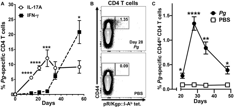
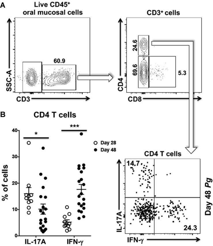
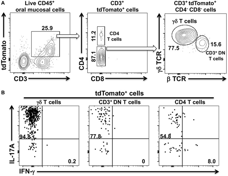
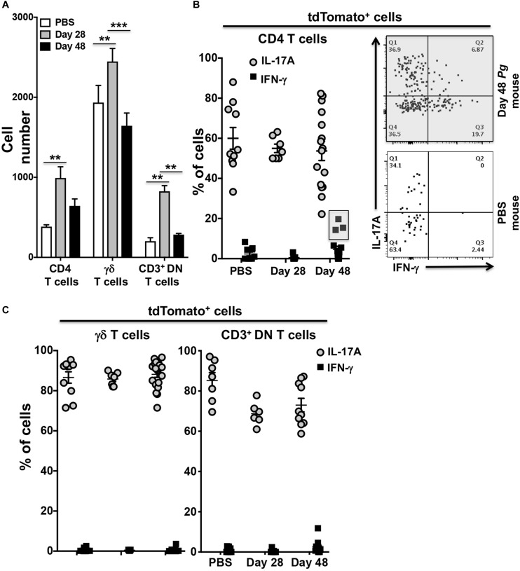
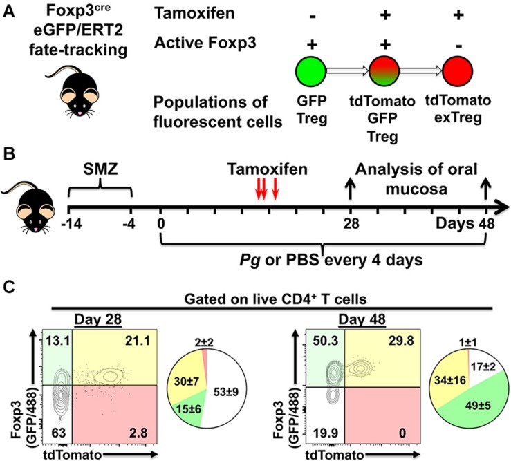
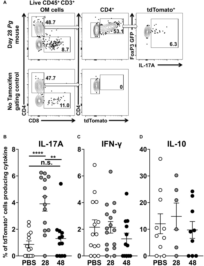
In periodontitis Porphyromonas gingivalis contributes to the development of a dysbiotic oral microbiome. This altered ecosystem elicits a diverse innate and adaptive immune response that simultaneously involves Th1, Th17, and Treg cells. It has been shown that Th17 cells can alter their gene expression to produce interferon-gamma (IFN-γ). Forkhead box P3 (Foxp3) is considered the master regulator of Treg cells that produce inhibitory cytokines like IL-10. Differentiation pathways that lead to Th17 and Treg cells from naïve progenitors are considered antagonistic. However, it has been reported that Treg cells expressing IL-17A as well as IFN-γ producing Th17 cells have been observed in several inflammatory conditions. Each scenario appears plausible with T cell transdifferentiation resulting from persistent microbial challenge and consequent inflammation. We established that oral colonization with P. gingivalis drives an initial IL-17A dominated Th17 response in the oral mucosa that is dependent on intraepithelial Langerhans cells (LCs). We hypothesized that Treg cells contribute to this initial IL-17A response through transient expression of IL-17A and that persistent mucosal colonization with P. gingivalis drives Th17 cells toward an IFN-γ phenotype at later stages of infection. We utilized fate-tracking mice where IL-17A- or Foxp3-promoter activity drives the permanent expression of red fluorescent protein tdTomato to test our hypothesis. At day 28 of infection timeline, Th17 cells dominated in the oral mucosa, outnumbering Th1 cells by 3:1. By day 48 this dominance was inverted with Th1 cells outnumbering Th17 cells by nearly 2:1. Tracking tdTomato+ Th17 cells revealed only sporadic transdifferentiation to an IFN-γ-producing phenotype by day 48; the appearance of Th1 cells at day 48 was due to a late de novo Th1 response. tdTomato+ Foxp3+ T cells were 35% of the total live CD4+T cells in the oral mucosa and 3.9% of them developed a transient IL-17A-producing phenotype by day 28. Interestingly, by day 48 these IL-17A-producing Foxp3+ T cells had disappeared. Therefore, persistent oral P. gingivalis infection stimulates an initial IL-17A-biased response led by Th17 cells and a small but significant number of IL-17A-expressing Treg cells that changes into a late de novo Th1 response with only sporadic transdifferentiation of Th17 cells.

Keywords: Foxp3; IL-17A; Porphyromonas gingivalis; Th17 cells; Treg cells; fate-tracking; periodontitis.


Figures

FIGURE 1
FIGURE 1
The percentage of P. gingivalis-specific CD4+ T cells expressing IFN-γ increases from day 28 in cervical lymph nodes. Single-cell suspensions from cervical lymph nodes of C57BL/6J mice orally inoculated with Pg or PBS were enriched for CD4+ cells at defined time points and stimulated with PMA/ionomycin in the presence of brefeldin A. Cells were surface stained with anti-mouse CD3, B220, CD8α, CD4, CD44 fluorochrome-conjugated mAbs and pR/Kgp:I-Ab tetramer and then intracellularly with anti-mouse IL-17A and IFN-γ mAbs to identify antigen-experienced gingipain-specific CD4+ T cells by flow cytometry (gated as CD44bright CD3+ CD4+ pR/Kgp-IAb+ B220 CD8α). Data was pooled from three independent experiments totaling at least 8 mice per group and displayed as mean percentage ± SEM. (A) Summary data of total antigen-experienced pR/Kgp:I-Ab tetramer positive CD4+ T cells that expressed either IL-17A or IFN-γ. (B) Representative FACS plots from a Pg and PBS mouse showing pR/Kgp:I-Ab tetramer positive CD4+ T cells. Gates are drawn around antigen-experienced CD4+ T cells identified as CD44bright. The frequency of pR/Kgp:I-Ab tetramer positive cells identified as a percentage of the total antigen-experienced CD4+ T cell population is given. (C) Summary data of percentage of antigen-experienced pR/Kgp:I-Ab tetramer positive CD4+ T cells. Percentages were compared using two-tailed Student’s t-test. *p < 0.05, **p < 0.01, ***p < 0.001, ****p < 0.0001.
FIGURE 2
FIGURE 2
Frequency of Th1 cells significantly increases within the oral mucosa of P. gingivalis inoculated mice by day 48. (A) Representative flow cytometry plots showing the gating strategy used to identify Th1 (IFN-γ+) and Th17 (IL-17A+) cells within the oral mucosa. Numbers alongside gates indicate% of cells within that gate. (B) Summary data showing individual data points from three experiments examining the frequency of Th1 and Th17 recovered from the oral mucosa of mice at day 28 and day 48. Means ± SEM are plotted. Groups were compared with two-tailed Student’s t-test. *p < 0.05, ***p < 0.001.
FIGURE 3
FIGURE 3
tdTomato expression in three distinct CD3+ T cells populations within the oral mucosa of IL-17Acre fate-tracking mice. Oral mucosa was harvested from IL-17Acre fate-tracking mice and single cell suspensions prepared from the tissue for subsequent analysis by flow cytometry. Single cell suspensions were stained with vitality dye Zombie Aqua followed by a panel of anti-mouse mAbs to identify immune cell subsets expressing tdTomato. Cells were counted by flow cytometry and analyzed by FlowJo software. (A) Representative flow cytometry plots showing gating strategy to identify three tdTomato+ cell populations. Numbers indicate the% of cells within a particular gate. Live CD4+ T cells were identified as Zombie Aqualo, CD45+, CD3+, CD4+, CD8α, NK1.1; live γδT cells as Zombie Aqualo, CD45+, CD3+, γδ TCR+, β TCR, CD4, CD8α, NK1.1; live CD3+ DN T cells as Zombie Aqualo, CD45+, CD3+, β TCR+, γδ TCR, CD4, CD8α, NK1.1. (B) Representative flow cytometry plots showing expression of IL-17A and IFN-γ in the three CD3+ tdTomato+ cell populations. Single cell suspensions obtained from oral mucosa were cultured and stimulated with PMA/ionomycin in the presence of brefeldin A. Cells were surface stained with anti-mouse mAbs as in (A) and then intracellularly with anti-mouse IL-17A and IFN-γ mAbs.
FIGURE 4
FIGURE 4
Expression of IFN-γ by tdTomato+ CD4+ T cells present within the oral mucosa of P. gingivalis inoculated mice is sporadic. IL-17Acre fate-tracking mice were pre-treated with SMZ and then orally inoculated with either P. gingivalis (4 × 10⁹ cfu per ml) or vehicle (PBS). At day 28 or 48 oral mucosa was harvested and single-cell suspensions from single mice treated and stained with mAbs as described in Figure 3. (A) Summary data of total numbers of CD4+ T cells, CD3+ DN T cells and γδ T cells found in the oral mucosa of mice after 28 or 48 days. Cell types were identified from single cell suspensions as described in Figure 3. Cell numbers were normalized to 100,000 live non-immune cells to account for potential cell loss during processing and counting. Data are from 3 experiments with at least 2 mice per time point and are plotted with means ± SEM. Means analyzed by two-tailed Student’s t-test. **p < 0.01, ***p < 0.001. (B) Summary data showing individual data points from three experiments examining the frequency of IL-17A and IFN-γ expression in IL-17Acre-tdTomato+ CD4+ T cells recovered from the oral mucosa of IL17A fate-tracking mice. Representative flow cytometry plots from a single PBS and P. gingivalis inoculated mouse at day 48 showing evidence of IFN-γ expression in IL-17Acre-tdTomato+ CD4+ T cells. (C) Summary data showing individual data points from three experiments examining the frequency of IL-17A and IFN- γ expression in IL-17Acre-tdTomato+ CD3+ DN T cells and γδ T cells recovered from the oral mucosa of IL-17Acre fate-tracking mice.
FIGURE 5
FIGURE 5
Experimental design to identify populations of fluorescently labeled Treg cells. (A) Potential populations of fluorescent Treg cells found in tamoxifen-treated Foxp3cre-GFP//ERT2 fate-tracking mice. (B) Experimental timeline for administration of P. gingivalis and, tamoxifen in experimental animals. (C) Representative flow cytometry plots gated on live CD4+ T cells from the oral mucosa of mice treated with tamoxifen and inoculated with Pg for 28 or 48 days. CD4+ T cells were identified as Zombie Aqualo, CD45+, CD3+, CD4+, β TCR+, CD8α. To augment GFP expressed from the Foxp3 promoter in these mice, cells were additionally stained with anti-mouse Foxp3 mAb conjugated to AF488. Numbers within the quadrants indicate% of the total CD4+ T cells gate. Summary pie charts show mean% ± SEM N = 4 mice.
FIGURE 6
FIGURE 6
P. gingivalis induces transient IL-17A expression in Treg cells in the oral mucosa. Single cell suspensions were isolated from oral mucosa of P. gingivalis inoculated mice at 28 or 48 days. Single cell suspensions were cultured and stimulated with PMA/ionomycin in the presence of brefeldin A. Cultured cells were surface stained with anti-mouse CD3, B220, CD8α, and CD4 fluorochrome-conjugated mAbs to identify tdTomato+ CD4+ Treg cells by flow cytometry (gated as CD3+ CD4+ tdTomato+ B220 CD8α) and then intracellular stained with anti-mouse IL-17A, IFN-γ, and IL-10. (A) Representative flow cytometry plots showing gating strategy to identify the Foxp3cre-tdTomato+ cell population within the oral mucosa also expressing Foxp3-GFP and IL-17A. Numbers indicate the % of cells within the associated gate. (B–D) Means of the frequency of tdTomato+ CD4+ T cells expressing IL-17A (B), IFN- γ (C), or IL-10 (D) at day 28 (gray circle) and 48 (black circle) were compared to sham controls (white circle) using two-tailed Student’s t-test and presented as means ± SEM. **p < 0.01, ****p < 0.0001. Each circle represents two pooled oral mucosae from two mice.
FIGURE 7
FIGURE 7
Foxp3cre-tdTomato+ Treg cells producing IL-17A have an active Foxp3 promoter. Foxp3cre-tdTomato+ CD4+ Treg cells from the oral mucosa were identified by flow cytometry (gated as CD3+ CD4+ tdTomato+ B220 CD8α). As the GFP signal in these mice was weak, anti-mouse FoxP3-Alexa488 was added during intracellular staining to increase the Foxp3 signal. The mean fluorescence intensity (MFI) of Foxp3-dependent green fluorescent protein (GFP) in populations of tdTomato (white bars) and tdTomato+ cells (black bars) from 20 mice are compared and presented for cells expressing IL-17A, IFN-γ or neither of the two cytokines. The GFP MFI was compared by fitting a mixed effects model with paired values. A post hoc Sidak’s multiple comparisons test was utilized to determine p-values of comparisons found significant by the initial mixed-effects analysis. *p < 0.05, ****p < 0.0001.

Similar articles

Cited by

References

    1. Kassebaum NJ, Bernabe E, Dahiya M, Bhandari B, Murray CJ, Marcenes W. Global burden of severe periodontitis in 1990-2010: a systematic review and meta-regression. J Dent Res. (2014) 93:1045–53. 10.1177/0022034514552491 - DOI - PMC - PubMed
    1. Eke PI, Thornton-Evans GO, Wei L, Borgnakke WS, Dye BA, Genco RJ. Periodontitis in US adults: national health and nutrition examination survey 2009-2014. J Am Dent Assoc. (2018) 149:576–88.e6. 10.1016/j.adaj.2018.04.023 - DOI - PMC - PubMed
    1. Neely AL, Holford TR, Loe H, Anerud A, Boysen H. The natural history of periodontal disease in humans: risk factors for tooth loss in caries-free subjects receiving no oral health care. J Clin Periodontol. (2005) 32:984–93. 10.1111/j.1600-051X.2005.00797.x - DOI - PubMed
    1. Costalonga M, Herzberg MC. The oral microbiome and the immunobiology of periodontal disease and caries. Immunol Lett. (2014) 162(2 Pt A):22–38. 10.1016/j.imlet.2014.08.017 - DOI - PMC - PubMed
    1. Lamont RJ, Koo H, Hajishengallis G. The oral microbiota: dynamic communities and host interactions. Nat Rev Microbiol. (2018) 16:745–59. 10.1038/s41579-018-0089-x - DOI - PMC - PubMed

Publication types

MeSH terms