

Ultrathin Descemet stripping automated endothelial keratoplasty versus Descemet membrane endothelial keratoplasty: a fellow-eye comparison
- PMID: 32391399
- PMCID: PMC7201595
- DOI: 10.1186/s40662-020-00191-6
Ultrathin Descemet stripping automated endothelial keratoplasty versus Descemet membrane endothelial keratoplasty: a fellow-eye comparison
Abstract
Background: To compare the visual outcome and patients' satisfaction after ultrathin Descemet stripping automated endothelial keratoplasty (UT-DSAEK) and Descemet membrane endothelial keratoplasty (DMEK) performed on fellow eyes of the same patients.
Methods: In this retrospective study, the records of 18 pseudophakic patients affected by Fuchs endothelial dystrophy who underwent DMEK in one eye and UT-DSAEK in the fellow eye were reviewed. Best corrected visual acuity (BCVA), corneal pachymetry, keratometry, corneal aberrations, photopic and mesopic contrast sensitivity, and endothelial cell counts measured 12 months after surgery in either eye were analyzed and compared. The results of a satisfaction questionnaire were also reviewed.
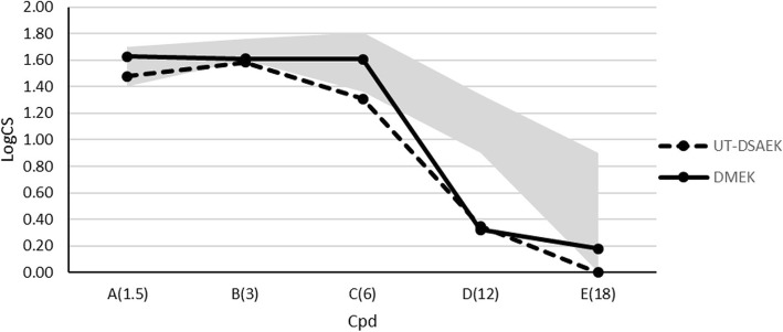
Results: Twelve months after surgery, BCVA was not significantly different in UT-DSAEK and DMEK eyes (0.10 ± 0.04 and 0.07 ± 0.07 logMAR, respectively); at both 4- and 6 mm optical zones total and posterior corneal higher order aberrations (HOAs), posterior astigmatism and total coma were significantly lower after DMEK; BCVA in both groups was significantly correlated mainly with anterior corneal aberrations; contrast sensitivity was higher after DMEK especially in mesopic conditions and at medium spatial frequencies; the endothelial cell density was similar, although slightly higher in the UT-DSAEK group (p = 0.10). The satisfaction questionnaire showed that although patients were highly satisfied from both procedures, more than half of them preferred DMEK and reported a more comfortable and quicker postoperative recovery.
Conclusions: DMEK and UT-DSAEK showed no evidence of difference in terms of postoperative BCVA, although DMEK had a better performance in terms of contrast sensitivity, posterior corneal aberrations and overall patient satisfaction.
Keywords: DMEK; DSAEK; Descemet membrane endothelial keratoplasty; Descemet stripping automated endothelial keratoplasty; UT-DSAEK; Ultra-thin Descemet stripping automated endothelial keratoplasty.
© The Author(s) 2020.
Conflict of interest statement
Competing interestsThe authors declare that they have no competing interests.
Figures

References
-
- Dickman MM, Kruit PJ, Remeijer L, van Rooij J, Van der Lelij A, Wijdh RH, et al. A randomized multicenter clinical trial of ultrathin Descemet stripping automated endothelial keratoplasty (DSAEK) versus DSAEK. Ophthalmology. 2016;123(11):2276–84. - PubMed
LinkOut - more resources
Full Text Sources
