. 2020 Apr 21;5(17):10077-10088.
doi: 10.1021/acsomega.0c00615. eCollection 2020 May 5.

cis-9-Hexadecenal, a Natural Compound Targeting Cell Wall Organization, Critical Growth Factor, and Virulence of Aspergillus fumigatus

Affiliations

cis-9-Hexadecenal, a Natural Compound Targeting Cell Wall Organization, Critical Growth Factor, and Virulence of Aspergillus fumigatus

Shanu Hoda et al. ACS Omega. .

Abstract

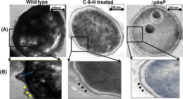
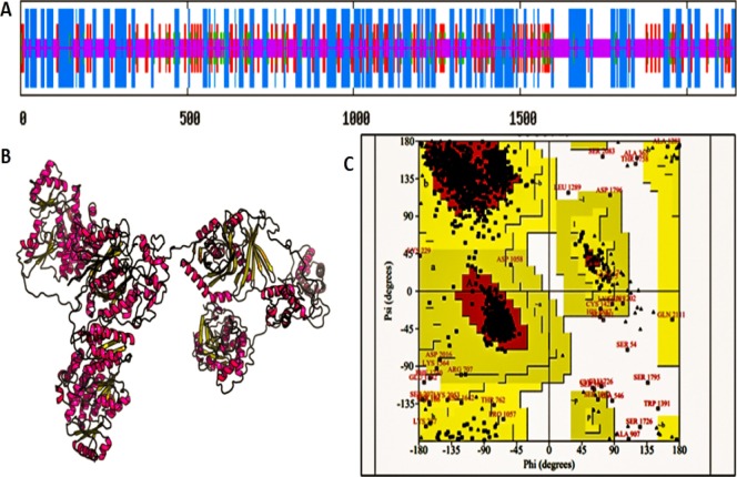
Aspergillus fumigatus causes several nosocomial pulmonary infections and accounts for high morbidity and mortality rate globally. Among various virulence factors, 1,8-dihydroxynaphthalene-melanin plays an important role in the survival during unfavorable conditions both in vivo and in vitro, masks various molecular patterns associated with A. fumigatus, and protects it from the host immune system. In the present study, we aim to understand the potential of cis-9-hexadecenal as an antimelanogenic compound and its role in modulating other associated virulence factors in A. fumigatus. cis-9-Hexadecenal is a bioactive compound that belongs to C16 mono-unsaturated fatty-aldehyde groups. Minimum effective concentration of cis-9-hexadecenal affecting A. fumigatus melanin biosynthesis was determined using broth microdilution method. The spectrophotometric analysis revealed reduced melanin content (91%) and hydrophobicity (59%) at 0.293 mM of cis-9-hexadecenal. Cell surface organizational changes using electron microscopy showed altered demelanized smooth A. fumigatus conidial surface without any protrusions after cis-9-hexadecenal treatment. The transcript analysis of polyketide synthase (PKS) pksP/alb1 gene was quantified through qRT-PCR which revealed an upregulated expression. Total proteome profiling conducted through LC-MS-MS showed upregulated PKS enzyme but other downstream proteins involved in the 1,8-dihydroxynaphthalene-melanin biosynthesis pathway were absent. The homology modeling of PKS using Expasy's web server predicted that PKS is stable at varied conditions and is hydrophilic in nature. The Ramachandran plot by PROCHECK confirmed the 3-D structure of PKS to be reliable. Docking analysis using AutoDock-4.2.6 predicted the binding of cis-9-hexadecenal and PKS at Thr-264 and Ser-171 residue via hydrogen bonding at a low binding energy of -4.95 kcal/mol.


Conflict of interest statement

The authors declare no competing financial interest.

Figures

Figure 1
Figure 1
Estimation of MEC of cis-9-hexadecenal (A), AmpB (B), and DMSO (C). Demelanized colonies were observed in C-9-H-treated wells at 0.293 mM concentration, whereas no demelanization was observed in AmpB- and DMSO-treated wells.
Figure 2
Figure 2
MEC of the compound cis-9-hexadecenal for analyzing antimelanogenic efficacy in (A) M. oryzae, (B) A. terreus, and (C) A. flavus. Demelanized colonies were observed only in M. oryzae at 2.34 mM concentration of C-9-H whereas no demelanization was observed in A. terreus and A. flavus.
Figure 3
Figure 3
Phenotype-based visual observation of A. fumigatus conidial color in the presence of DHN-melanin inhibitors (TC and PQ), phytocompound C-9-H, and antifungal drug AmpB. A. fumigatus was cultured on CzA supplemented with TC, PQ, AmpB, and C-9-H. The color of the colonies was observed visually and compared with that of WT and ΔpksP. WT—wild type, TC—tricyclazole, PQ—pyroquilon, C-9-H—cis-9-hexadecenal, AmpB—amphotericin B, ΔpksPpksP mutant.
Figure 4
Figure 4
Evaluation of the physical properties of the conidial surface. The WT A. fumigatus without any treatment (positive control), WT with C-9-H (0.293 mM), AmpB (0.012 mM; drug control) and ΔpksP strain (negative control) were grown on Czapek medium (n = 3). (A) Melanin estimation in treated and control group (p ≤ 0.0087), (B) UV–vis spectrum of melanin showing a characteristic peak at UV-region 200–260 with gradual decrease in absorption toward visible range where red, black, and blue curve shows the melanin spectra of WT, AmpB, and C-9-H respectively, and (C) altered membrane surface depicted by reduced cell surface hydrophobicity (CSH) (p ≤ 0.0071).
Figure 5
Figure 5
Visualization of the A. fumigatus conidial surface by SEM. Magnification is 50k× and scale corresponding to 200 nm. WT—wild type, C-9-H—cis-9-hexadecenal-treated, AmpB—amphotericin B–treated, and ΔpksPpksP mutant A. fumigatus.
Figure 6
Figure 6
Ultrastructure of the lateral section of wild type (WT), cis-9-hexadecenal (C-9-H)–treated, and pksP mutant (ΔpksP) A. fumigatus conidia visualized by TEM. C-9-H and ΔpksP have a smooth surface (black arrowheads), whereas WT have a rough sough surface with melanin deposition (yellow arrowheads) and protrusions (blue arrow). Magnifications (A) 10k×, (B) 20k×.
Figure 7
Figure 7
Relative expression fold of pksP/alb1 gene with wild type (WT), cis-9-hexadecenal (C-9-H) treated, and pksP mutant (ΔpksP) A. fumigatus (p ≤ 0.0005). RNA was extracted from 4 days old untreated WT, C-9-H treated, and (ΔpksP) A. fumigatus (n = 3). β-Actin expression was used as an internal control. mRNA expression corresponding to the pksp/alb1 gene was compared with that expressed in the untreated A. fumigatus.
Figure 8
Figure 8
GO study based on (A) cellular and (B) biological functions. On the basis of cellular localization 36% membrane proteins and 7% of extracellular proteins were differentially expressed in WT- and C-9-H-treated sample. Biological GO showed that 3% cell wall integrity proteins, 9% secondary metabolites, and 5% cell stress proteins were differentially expressed between proteins isolated from WT- and C-9-H-treated A. fumigatus respectively.
Figure 9
Figure 9
(A) Secondary structure of PKS protein of A. fumigatus as predicted by SOPMA software. SOPMA: Self Optimized Prediction Method with Alignment. α-Helixes are depicted by blue lines, β-sheets by red lines, turns by green lines and coils by purple lines, (B) tertiary structure of PKS protein of A. fumigatus as predicted by RaptorX software, (C) Ramachandran plot of predicted 3-D model of PKS protein of A. fumigatus by PROCHECK software. The bottom left box indicates the presence of right-handed α-helix. The red region indicates the favored region where no steric clashes occur. The majority of amino acids are present in phi–psi distribution.
Figure 10
Figure 10
Docking study of the compounds with the active domain of A. fumigatus PKS protein. Protein–ligand interaction was visualized by UCSF Chimera. The bond between PKS and C-9-H is marked with blue arrow. The amino acid residues (SER-171 and THR 264) forming bonds are marked with blue circles. Green arrow shows the ligand C-9-H.

References

    1. Bhetariya P. J.; Madan T.; Basir S. F.; Varma A.; Usha S. P. Allergens/antigens, toxins and polyketides of important Aspergillus species. Indian J. Clin. Biochem. 2011, 26, 104–119. 10.1007/s12291-011-0131-5. - DOI - PMC - PubMed
    1. van de Veerdonk F. L.; Gresnigt M. S.; Romani L.; Netea M. G.; Latgé J.-P. Aspergillus fumigatus morphology and dynamic host interactions. Nat. Rev. Microbiol. 2017, 15, 661–674. 10.1038/nrmicro.2017.90. - DOI - PubMed
    1. Scorzoni L.; de Paula e Silva A. C. A.; Marcos C. M.; Assato P. A.; de Melo W. C. M. A.; de Oliveira H. C.; Costa-Orlandi C. B.; Mendes-Giannini M. J. S.; Fusco-Almeida A. M. Antifungal therapy: new advances in the understanding and treatment of mycosis. Front. Microbiol. 2017, 8, 36.10.3389/fmicb.2017.00036. - DOI - PMC - PubMed
    1. Joly V.; Saint-Pierre-Chazalet M.; Saint-Julien L.; Bolard J.; Carbon C.; Yeni P. Inhibiting cholesterol synthesis reduces the binding and toxicity of amphotericin B against rabbit renal tubular cells in primary culture. J. Infect. Dis. 1992, 165, 337–343. 10.1093/infdis/165.2.337. - DOI - PubMed
    1. Boominathan M.; Ramamurthy V. Antimicrobial activity of Heliotropium indicum and Coldenia procumbens. J. Ecobiol. 2009, 24, 11–15.