. 2020 Aug;103(4):1399-1419.
doi: 10.1111/tpj.14808. Epub 2020 Jun 12.

Exocyst mutants suppress pollen tube growth and cell wall structural defects of hydroxyproline O-arabinosyltransferase mutants

Affiliations

Exocyst mutants suppress pollen tube growth and cell wall structural defects of hydroxyproline O-arabinosyltransferase mutants

Steven Beuder et al. Plant J. 2020 Aug.

Abstract

HYDROXYPROLINE O-ARABINOSYLTRANSFERASEs (HPATs) initiate a post-translational protein modification (Hyp-Ara) found abundantly on cell wall structural proteins. In Arabidopsis thaliana, HPAT1 and HPAT3 are redundantly required for full pollen fertility. In addition to the lack of Hyp-Ara in hpat1/3 pollen tubes (PTs), we also found broadly disrupted cell wall polymer distributions, particularly the conversion of the tip cell wall to a more shaft-like state. Mutant PTs were slow growing and prone to rupture and morphological irregularities. In a forward mutagenesis screen for suppressors of the hpat1/3 low seed-set phenotype, we identified a missense mutation in exo70a2, a predicted member of the vesicle-tethering exocyst complex. The suppressed pollen had increased fertility, fewer morphological defects and partially rescued cell wall organization. A transcriptional null allele of exo70a2 also suppressed the hpat1/3 fertility phenotype, as did mutants of core exocyst complex member sec15a, indicating that reduced exocyst function bypassed the PT requirement for Hyp-Ara. In a wild-type background, exo70a2 reduced male transmission efficiency, lowered pollen germination frequency and slowed PT elongation. EXO70A2 also localized to the PT tip plasma membrane, consistent with a role in exocyst-mediated secretion. To monitor the trafficking of Hyp-Ara modified proteins, we generated an HPAT-targeted fluorescent secretion reporter. Reporter secretion was partially dependent on EXO70A2 and was significantly increased in hpat1/3 PTs compared with the wild type, but was reduced in the suppressed exo70a2 hpat1/3 tubes.

Keywords: Arabidopsis thaliana; cell wall; exocyst; glycoprotein; pollen tube; secretion; tip growth.


Conflict of interest statement

The authors declare no conflicts of interest.

Figures
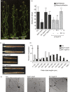
Figure 1
Figure 1
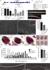
Suppressor mutant frh1 increases the pollen fertility of hpat1 hpat3 plants. (a) Both double mutant hpat1/3 plants and the suppressed triple mutant frh1 hpat1/3 plants appeared morphologically normal, aside from differences in seed set. (b) Average number of seeds per silique (±SD, n ≥ 10 per genotype) for the indicated genotypes. The low‐seed‐set phenotype of hpat1/3 double mutants is suppressed by frh1. High seed set is also observed for heterozygous frh1 plants. Reciprocal crosses between hpat1/3 and frh1 hpat1/3 plants demonstrated that frh1 suppression occurs in the pollen. ***Statistically significant difference (Student’s t‐test, P < 0.0005) compared with the hpat1/3 value for either the self‐fertilized siliques or the manually pollinated samples. NS is not significantly different. (c) Cleared siliques of wild‐type (WT) Columbia, hpat1 hpat3 and frh1 hpat1/3 plants. WT and frh1 hpat1/3 images are composite images to allow full silique imaging. (d) Histogram of distribution of pollen tube (PT) lengths after 5 h of in vitro growth for the indicated genotypes. Data for all genotypes are statistically significantly different from both other genotypes (Student’s t‐test, P < 0.005). (e) In vitro grown PTs of the indicated genotypes. Scale bars: 100 μm.
Figure 2
Figure 2
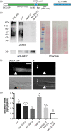
frh1 partially suppresses the disrupted polarity of cell wall polymers in hpat1/3 pollen tubes (PTs). (a) Wild‐type (WT) Columbia‐0 PT stained with JIM20 primary antibody and anti‐rat FITC‐conjugated secondary antibody. (b) Left: Maximum projections of PTs stained with aniline blue fluorochrome (ABF). Right: quantification of signal intensity from the tip (distance on x‐axis = 0) to 50 μm down the shaft of the PT (for more details concerning image analysis, see Experimental procedures). Colored lines represent the mean fluorescent intensities for each genotype, and shading represents the standard error. n ≥ 30 for each genotype. The vertical dashed line represents the approximate region of the PT where the apical dome transitions to the shaft. (c) Left: medial Z‐slices of PTs stained with LM20 primary antibody and anti‐rat FITC‐conjugated secondary antibody. Right: quantification of fluorescence intensity as in (b), except measurements were taken to 25 μm from the tip. (d) Left: medial Z‐slice of PTs stained with LM19 primary antibody and anti‐rat FITC‐conjugated secondary antibody. Right: quantification of signal intensity as in (c). All images were acquired by confocal microscopy at 100× magnification. Scale bars: 10 μm.
Figure 3
Figure 3
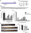
EXO70A2 is required for efficient pollen germination and pollen tube (PT) growth. (a) Diagram of the EXO70A2 coding sequence, with the position of the exo70a2‐2 (G319E) missense mutation and insertion mutants exo70a2‐D (Hála et al., 2008) and exo70a2‐3 marked. (b) Semi‐quantitative RT‐PCR of flower cDNA samples from the indicated genotypes showing an absence of transcript in the exo70a2‐3 plants using the primer pairs indicated in (a). (c) Average number of seeds per silique (±SD) for plants of the indicated genotype. Note the modest reduction in seed set for exo70a2‐3 in the wild‐type (WT) background along with the partial suppression of the hpat1 hpat3 phenotype by this mutation. ***Statistically significant differences with the corresponding background (Columbia or hpat1 hpat3, Student’s t‐test, P < 0.0005, n ≥ 12). (d) Cleared siliques of the exo70a2 mutant alleles in the Columbia background, see Figure 1(d) for Col comparison image. (e and f) In vitro pollen germination samples of Columbia (e) and exo70a2‐3 (f) 3 h after transfer to growth media. Inset in (f) shows non‐germinated exo70a2‐3 pollen grains. (g) Quantification of pollen germination frequencies for WT, exo70a2‐2 and exo70a2‐3. Mean of three replicates of ≥740 pollen grains, ±SD. **Significant difference between WT (Student’s t‐test, P < 0.005). (h–j) Alexander viability staining of anthers of WT (h), exo70a2‐2 (i) and exo70a2‐3 (j). Insets show free pollen grains. Note no difference in viability staining between genotypes. (k and l) Pollen grains stained with Ruthenium red to mark pectin accumulation at the germination plaque. (m) Histogram of PT lengths after 5 h of in vitro growth for the indicated genotypes. Data for all genotypes are statistically significantly different from both other genotypes (Student’s t‐test, P < 0.005, n ≥ 200 per genotype). (n) sustained growth rate of PTs of the indicated genotype. Mean, n ≥ 21 tubes, ±SD. (o and p) Differential interference contrast (DIC) micrograph of PTs.
Figure 4
Figure 4
EXO70A2 localizes to the tip of growing pollen tubes (PTs). Representative PT expressing EXO70A2:mNG under its native promoter and co‐stained with FM4‐64 to visualize the plasma membrane. Single channels and merged image shown; overlapping signal is false‐colored white. Imaged with confocal microscopy at 100× magnification. Scale bar: 10 μm.
Figure 5
Figure 5
Secretion of GF(EXT3)P is decreased in hpat1/3 exo70a2‐2 pollen tubes (PTs). (a) Schematic of the GF(EXT3)P construct, which includes the EXT3 signal peptide (SP), amino acids 1–175 of GFP, an 8× HIS tag, a portion of EXT3, a Myc tag, and amino acids 176–241 of GFP. (b) Western blots (left) and corresponding Ponceau‐stained membrane (right) of PT protein samples. *Samples from plants carrying the LAT52:GF(EXT3)P transgene; NT, non‐transgenic. An anti‐GFP polyclonal antibody detects the fusion protein in the transgenic lines at the expected mass (~34 kDa). The reporter is also detected by the Hyp‐Ara monocolonal antibody JIM20 only in the transgenic Col sample. (c) Images of plasmolyzed PTs expressing LAT52:GF(EXT3)P (left) and non‐transgenic control (right). Scale barL 10 μm. Arrows indicate the location of the PT cell wall tip, and arrowheads mark the plasma membrane. (d) Quantification of secreted GFP signal reported as a secretion index (SI: mean ± SE, n ≥ 30 PTs per genotype; for details, see Experimental procedures). Samples labeled with the same letter are not significantly different (Benjamini–Hochberg flase‐discovery rate, FDR ≤ 0.05).
Figure 6
Figure 6
Mutations in exocyst complex member sec15a also suppress the hpat1/3 fertility phenotype. (a) Gene model diagram showing the relative position of insertion in the sec15a‐2 allele and the suppressor allele identified in frh2 (sec15a‐3). (b) Segregation of the sec15a‐3 mutation in the frh2 hpat1/3 BC5 F2 population. As expected for an hpat1/3‐suppressing mutation, the number of homozygous wild‐type SEC15A plants identified (+/+) was significantly below the expected value based on Mendelian segregation (chi‐square test, P = 1.57 × 10−5). (c) Sample sec15a‐3 genotyping data for 15 frh2 hpat1/3 BC5 F2 individuals visualized on an agarose gel. The wild‐type (WT) allele is cleaved by digestion with MnlI. The single homozygous WT individual marked by ‘*’ did not have the suppressive phenotype. (d) Average number of seeds per silique (±SD) for plants of the indicated genotypes. sec15a‐2 is not transmitted through the male and is not recoverable as a homozygous mutant (Hála et al., 2008). Both sec15a alleles suppress the hpat1/3 phenotype, with homozygous mutants showing stronger suppression than heterozygotes. ‘ns’ marks samples that were not statistically different from their corresponding background genotype. ***Statistically different samples (Student’s t‐test, P < 0.0005, n ≥ 11). (e) Histogram of PT lengths after 5 h of in vitro growth for the indicated genotypes. Data for all genotypes are statistically significantly different from both other genotypes (Student’s t‐test, P < 0.005, n ≥ 100 per genotype). (f) Cleared siliques of the indicated genotypes. (g) Average number of seeds per silique (±SDs) for the indicated genotypes. All hpat1/3‐based genotypes were siblings segregating from the same F2 population. Statistically different samples (Student’s t‐test): ***P < 0.0005; *P < 0.05 (n ≥ 15).

References

    1. Adams, E. and Frank, L. (1980) Metabolism of proline and the hydroxyprolines. Annu Rev Biochem. 49, 1005–1061. - PubMed
    1. Alonso, J.M. , Stepanova, A.N. , Leisse, T.J. et al . (2003) Genome‐wide insertional mutagenesis of Arabidopsis thaliana. Science, 301, 653–657. - PubMed
    1. Bate, N. and Twell, D. (1998) Functional architecture of a late pollen promoter: pollen‐specific transcription is developmentally regulated by multiple stage‐specific and co‐dependent activator elements. Plant Mol. Biol. 37, 859–869. - PubMed
    1. Beuder, S. and MacAlister, C.A. (2020) Isolation and cloning of suppressor mutants with improved pollen fertility (2020) In: Pollen and Pollen Tube Biology: Methods and Protocols (Geitmann A., ed). MIMB, Humana Press. - PubMed
    1. Bloch, D. , Pleskot, R. , Pejchar, P. , Potocký, M. , Trpkošová, P. , Cwiklik, L. , Vukašinović, N. , Sternberg, H. , Yalovsky, S. and Žárský, V. (2016) Exocyst SEC3 and phosphoinositides define sites of exocytosis in pollen tube initiation and growth. Plant Physiol. 172, 980–1002. - PMC - PubMed

Publication types

MeSH terms