Combined CCNE1 high-level amplification and overexpression is associated with unfavourable outcome in tubo-ovarian high-grade serous carcinoma
- PMID: 32391646
- PMCID: PMC7578325
- DOI: 10.1002/cjp2.168
Combined CCNE1 high-level amplification and overexpression is associated with unfavourable outcome in tubo-ovarian high-grade serous carcinoma
Abstract
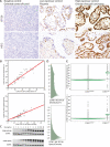
CCNE1 amplification is a recurrent alteration associated with unfavourable outcome in tubo-ovarian high-grade serous carcinoma (HGSC). We aimed to investigate whether immunohistochemistry (IHC) can be used to identify CCNE1 amplification status and to validate whether CCNE1 high-level amplification and overexpression are prognostic in HGSC. A testing set of 528 HGSC samples stained with two optimised IHC assays (clones EP126 and HE12) was subjected to digital image analysis and visual scoring. DNA and RNA chromogenic in situ hybridisation for CCNE1 were performed. IHC cut-off was determined by receiver operating characteristics (ROC). Survival analyses (endpoint ovarian cancer specific survival) were performed and validated in an independent validation set of 764 HGSC. Finally, combined amplification/expression status was evaluated in cases with complete data (n = 1114). CCNE1 high-level amplification was present in 11.2% of patients in the testing set and 10.2% in the combined cohort. The optimal cut-off for IHC to predict CCNE1 high-level amplification was 60% positive tumour cells with at least 5% strong staining cells (sensitivity 81.6%, specificity 77.4%). CCNE1 high-level amplification and overexpression were associated with survival in the testing and validation set. Combined CCNE1 high-level amplification and overexpression was present in 8.3% of patients, mutually exclusive to germline BRCA1/2 mutation and significantly associated with a higher risk of death in multivariate analysis adjusted for age, stage and cohort (hazard ratio = 1.78, 95 CI% 1.38-2.26, p < 0.0001). CCNE1 high-level amplification combined with overexpression identifies patients with a sufficiently poor prognosis that treatment alternatives are urgently needed. Given that this combination is mutually exclusive to BRCA1/2 germline mutations, a predictive marker for PARP inhibition, CCNE1 high-level amplification combined with overexpression may serve as a negative predictive test for sensitivity to PARP inhibitors.
Keywords: CCNE1; PARP inhibitor; amplification; cyclin E1; high grade serous carcinoma; ovarian cancer; prognosis.
© 2020 The Authors. The Journal of Pathology: Clinical Research published by The Pathological Society of Great Britain and Ireland & John Wiley & Sons Ltd.
Figures


References
Publication types
MeSH terms
Substances
Grants and funding
LinkOut - more resources
Full Text Sources
Medical
Miscellaneous

