[CT imaging features of patients with different clinical types of COVID-19]
- PMID: 32391664
- PMCID: PMC10412947
- DOI: 10.3785/j.issn.1008-9292.2020.03.05
[CT imaging features of patients with different clinical types of COVID-19]
Abstract
Objective: To investigate the CT findings of patients with different clinical types of coronavirus disease 2019 (COVID-19).
Methods: A total of 67 patients diagnosed as COVID-19 by nucleic acid testing were collected and divided into 4 groups according to the clinical stages based on Diagnosis and treatment of novel coronavirus pneumonia (trial version 6). The CT imaging characteristics were analyzed among patients with different clinical types.
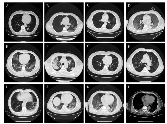
Results: Among 67 patients, 3(4.5%) were mild, 35 (52.2%) were moderate, 22 (32.8%) were severe, and 7(10.4%) were critical ill. No significant abnormality in chest CT imaging in mild patients. The 35 cases of moderate type included 3 (8.6%) single lesions, the 22 cases of severe cases included 1 (4.5%) single lesion and the rest cases were with multiple lesions. CT images of moderate patients were mainly manifested by solid plaque shadow and halo sign (18/35, 51.4%); while fibrous strip shadow with ground glass shadow was more frequent in severe cases (7/22, 31.8%). Consolidation shadow as the main lesion was observed in 7 cases, and all of them were severe or critical ill patients.
Conclusions: CT images of patients with different clinical types of COVID-19 have characteristic manifestations, and solid shadow may predict severe and critical illness.
目的: 探讨2019冠状病毒病(COVID-19)不同临床分型患者的胸部CT影像学特征。
方法: 收集经核酸检测确诊为COVID-19的患者67例,根据《新型冠状病毒感染肺炎诊疗方案(试行第六版)》对患者进行临床分型,其中轻型3例,普通型35例,重型22例,危重型7例。分析和比较不同临床分型患者的胸部CT影像学特征。
结果: 轻型患者胸部CT影像学检查无明显异常。普通型患者中,单发病灶3例(8.6%),多发病灶32例(91.4%);重型患者中,单发病灶1例(4.5%),多发病灶21例(95.5%);危重型患者均为多发病灶。普通型患者胸部CT影像以实性斑片影伴晕征为主要表现(18/35,51.4%);重型患者以条索影伴磨玻璃影为主要表现(7/22,31.8%);以实变影为主要表现共7例,全部为重型或危重型患者。
结论: COVID-19不同临床分型患者胸部CT影像学有特征性表现,以实变影为主要病灶的影像学特征可作为重型和危重型患者的指征之一。
References
-
- 中华人民共和国国家卫生健康委员会办公厅, 国家中医药管理局办公室.新型冠状病毒感染的肺炎诊疗方案(试行第六版)[A/OL].国卫办医涵[2020] 145号.(2020-02-18)[2020-02-28].. http://www.nhc.gov.cn/yzygj/s7653p/202002/8334a8326dd94d329df351d7da8aef...
-
- AI T, YANG Z L, ZHAN H Y, et al. Correlation of chest CT and RT-PCR testing in coronavirus disease 2019(COVID-19) in China:a report of 1014 cases. Radiology. 2020 doi: 10.1148/radiol.2020200642. [AI T, YANG Z L, ZHAN H Y, et al. Correlation of chest CT and RT-PCR testing in coronavirus disease 2019(COVID-19) in China:a report of 1014 cases[J]. Radiology, 2020. DOI:10.1148/radiol.2020200642.] - DOI - PMC - PubMed
-
- 中华医学会放射学分会 新型冠状病毒肺炎的放射学诊断:中华医学会放射学分会专家推荐意见(第一版) 中华放射学杂志. 2020;54 doi: 10.3760/cma.j.issn.1005-1201.2020.0001. [中华医学会放射学分会.新型冠状病毒肺炎的放射学诊断:中华医学会放射学分会专家推荐意见(第一版)[J].中华放射学杂志, 2020, 54. DOI:10.3760/cma.j.issn.1005-1201.2020.0001.] - DOI
-
- KOO H J, LIM S, CHOE J, et al. Radiographic and CT features of viral pneumonia. Radiographics. 2018;38(3):719–739. doi: 10.1148/rg.2018170048. [KOO H J, LIM S, CHOE J, et al. Radiographic and CT features of viral pneumonia[J]. Radiographics, 2018, 38(3):719-739. DOI:10.1148/rg.2018170048] - DOI - PubMed