

Induction of High Endothelial Venule-like Vessels in Oral and Cutaneous Lichen Planus: A Comparative Study
- PMID: 32391737
- PMCID: PMC7226624
- DOI: 10.1369/0022155420923272
Induction of High Endothelial Venule-like Vessels in Oral and Cutaneous Lichen Planus: A Comparative Study
Abstract
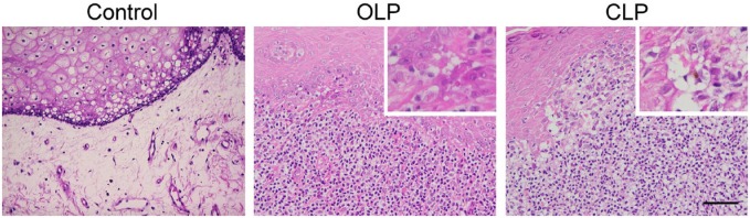
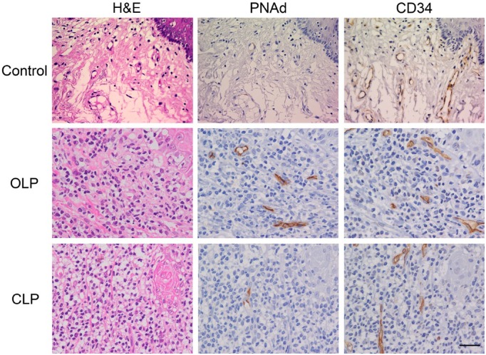
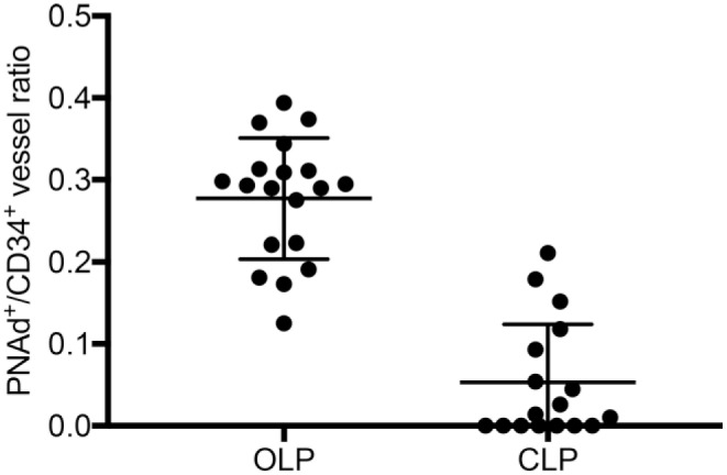
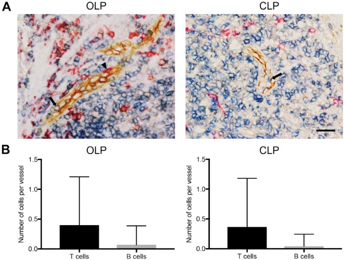
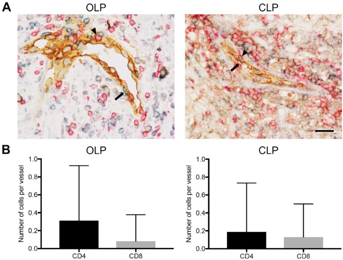
Lichen planus (LP) is a chronic inflammatory mucocutaneous disease involving the oral mucosa and skin. Both oral LP (OLP) and cutaneous LP (CLP) are histopathologically characterized by dense subepithelial lymphocyte infiltrates; however, the mechanisms underlying lymphocyte recruitment to sites of LP lesions are not fully understood. Here, we assessed the induction of peripheral lymph node addressin (PNAd)-expressing high endothelial venule (HEV)-like vessels in 19 OLP and 17 CLP cases. To do so, we performed immunohistochemical staining for PNAd and CD34, followed by quantitative analysis. We also conducted triple immunohistochemistry for PNAd and either CD3 and CD20 or CD4 and CD8 to identify the lymphocyte subset preferentially recruited via HEV-like vessels. PNAd-expressing HEV-like vessels were induced in and around lymphocyte aggregates in all cases of OLP and in 10 of 17 CLP cases, and these vessels were more frequently observed in OLP relative to CLP. Although the number of T-cells attached per HEV-like vessel exceeded the number of B-cells in both OLP and CLP, the number of CD4+ T-cells attached was greater than the number of CD8+ T-cells only in OLP. These findings combined suggest that PNAd-expressing HEV-like vessels play a more important role in the pathogenesis of OLP compared with CLP.
Keywords: chronic inflammation; high endothelial venule (HEV); lichen planus; lymphocyte recruitment; peripheral lymph node addressin (PNAd).
Conflict of interest statement
Figures
References
-
- Boyd AS, Neldner KH. Lichen planus. J Am Acad Dermatol. 1991;25(4):593–619. - PubMed
-
- Wagner G, Rose C, Sachse MM. Clinical variants of lichen planus. J Dtsch Dermatol Ges. 2013;11(4):309–19. - PubMed
-
- Weber B, Schlapbach C, Stuck M, Simon HU, Borradori L, Beltraminelli H, Simon D. Distinct interferon-gamma and interleukin-9 expression in cutaneous and oral lichen planus. J Eur Acad Dermatol Venereol. 2017;31(5):880–6. - PubMed
-
- Carbone M, Arduino PG, Carrozzo M, Gandolfo S, Argiolas MR, Bertolusso G, Conrotto D, Pentenero M, Broccoletti R. Course of oral lichen planus: a retrospective study of 808 northern Italian patients. Oral Dis. 2009;15(3):235–43. - PubMed
-
- Lage D, Pimentel VN, Soares TC, Souza EM, Metze K, Cintra ML. Perforin and granzyme B expression in oral and cutaneous lichen planus—a comparative study. J Cutan Pathol. 2011;38(12):973–8. - PubMed
Publication types
MeSH terms
Substances
LinkOut - more resources
Full Text Sources
Research Materials
Miscellaneous
