. 2020 Oct;297(1):E207-E215.
doi: 10.1148/radiol.2020201908. Epub 2020 May 11.

Abdominal Imaging Findings in COVID-19: Preliminary Observations

Affiliations

Abdominal Imaging Findings in COVID-19: Preliminary Observations

Rajesh Bhayana et al. Radiology. 2020 Oct.

Abstract

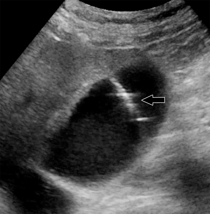
Background Angiotensin-converting enzyme 2, a target of severe acute respiratory syndrome coronavirus 2 (SARS-CoV-2), demonstrates its highest surface expression in the lung, small bowel, and vasculature, suggesting abdominal viscera may be susceptible to injury. Purpose To report abdominal imaging findings in patients with coronavirus disease 2019. Materials and Methods In this retrospective cross-sectional study, patients consecutively admitted to a single quaternary care center from March 27 to April 10, 2020, who tested positive for SARS-CoV-2 were included. Abdominal imaging studies performed in these patients were reviewed, and salient findings were recorded. Medical records were reviewed for clinical data. Univariable analysis and logistic regression were performed. Results A total of 412 patients (average age, 57 years; range, 18 to >90 years; 241 men, 171 women) were evaluated. A total of 224 abdominal imaging studies were performed (radiography, n = 137; US, n = 44; CT, n = 42; MRI, n = 1) in 134 patients (33%). Abdominal imaging was associated with age (odds ratio [OR], 1.03 per year of increase; P = .001) and intensive care unit (ICU) admission (OR, 17.3; P < .001). Bowel-wall abnormalities were seen on 31% of CT images (13 of 42) and were associated with ICU admission (OR, 15.5; P = .01). Bowel findings included pneumatosis or portal venous gas, seen on 20% of CT images obtained in patients in the ICU (four of 20). Surgical correlation (n = 4) revealed unusual yellow discoloration of the bowel (n = 3) and bowel infarction (n = 2). Pathologic findings revealed ischemic enteritis with patchy necrosis and fibrin thrombi in arterioles (n = 2). Right upper quadrant US examinations were mostly performed because of liver laboratory findings (87%, 32 of 37), and 54% (20 of 37) revealed a dilated sludge-filled gallbladder, suggestive of bile stasis. Patients with a cholecystostomy tube placed (n = 4) had negative bacterial cultures. Conclusion Bowel abnormalities and gallbladder bile stasis were common findings on abdominal images of patients with coronavirus disease 2019. Patients who underwent laparotomy often had ischemia, possibly due to small-vessel thrombosis. © RSNA, 2020.


Figures

(a) Axial and (b) coronal CT images of the abdomen and pelvis with intravenous contrast material in a 57-year-old man with high clinical suspicion for bowel ischemia. There is generalized small-bowel distension and segmental thickening (white arrows), with adjacent mesenteric congestion (black arrow) and a small volume of ascites (*). Findings are nonspecific but suggestive of early ischemia or infection.
Figure 1a:
(a) Axial and (b) coronal CT images of the abdomen and pelvis with intravenous contrast material in a 57-year-old man with high clinical suspicion for bowel ischemia. There is generalized small-bowel distension and segmental thickening (white arrows), with adjacent mesenteric congestion (black arrow) and a small volume of ascites (*). Findings are nonspecific but suggestive of early ischemia or infection.
Figure 1b:
(a) Axial and (b) coronal CT images of the abdomen and pelvis with intravenous contrast material in a 57-year-old man with high clinical suspicion for bowel ischemia. There is generalized small-bowel distension and segmental thickening (white arrows), with adjacent mesenteric congestion (black arrow) and a small volume of ascites (*). Findings are nonspecific but suggestive of early ischemia or infection.
(a) Coronal CT image of the abdomen and pelvis with intravenous contrast material in a 47-year-old man with abdominal tenderness shows typical findings of mesenteric ischemia and infarction, including pneumatosis intestinalis (white arrow) and nonenhancing bowel (*). Frank discontinuity of a thickened loop of small bowel in the pelvis (black arrow) is in keeping with perforation. (b) These findings are confirmed at laparotomy, with the additional observation of an atypical yellow discoloration of the bowel.
Figure 2a:
(a) Coronal CT image of the abdomen and pelvis with intravenous contrast material in a 47-year-old man with abdominal tenderness shows typical findings of mesenteric ischemia and infarction, including pneumatosis intestinalis (white arrow) and nonenhancing bowel (*). Frank discontinuity of a thickened loop of small bowel in the pelvis (black arrow) is in keeping with perforation. (b) These findings are confirmed at laparotomy, with the additional observation of an atypical yellow discoloration of the bowel.
Figure 2b:
(a) Coronal CT image of the abdomen and pelvis with intravenous contrast material in a 47-year-old man with abdominal tenderness shows typical findings of mesenteric ischemia and infarction, including pneumatosis intestinalis (white arrow) and nonenhancing bowel (*). Frank discontinuity of a thickened loop of small bowel in the pelvis (black arrow) is in keeping with perforation. (b) These findings are confirmed at laparotomy, with the additional observation of an atypical yellow discoloration of the bowel.
(a) Abdominal radiograph in a 52-year-old man show portal venous gas (arrow), suggestive of bowel infarction. (b) Postoperative CT image also shows portal venous gas (arrow). At laparotomy, bowel ischemia and necrosis are identified, along with an atypical yellow discoloration of the small bowel. (c) Photomicrograph shows submucosal arterioles with fibrin thrombi (arrowheads). The overlying mucosa (upper right) is partially necrotic, with crypt dropout and partial loss of the surface epithelium. (Hematoxylin-eosin stain; original magnification, ×400.)
Figure 3a:
(a) Abdominal radiograph in a 52-year-old man show portal venous gas (arrow), suggestive of bowel infarction. (b) Postoperative CT image also shows portal venous gas (arrow). At laparotomy, bowel ischemia and necrosis are identified, along with an atypical yellow discoloration of the small bowel. (c) Photomicrograph shows submucosal arterioles with fibrin thrombi (arrowheads). The overlying mucosa (upper right) is partially necrotic, with crypt dropout and partial loss of the surface epithelium. (Hematoxylin-eosin stain; original magnification, ×400.)
Figure 3b:
(a) Abdominal radiograph in a 52-year-old man show portal venous gas (arrow), suggestive of bowel infarction. (b) Postoperative CT image also shows portal venous gas (arrow). At laparotomy, bowel ischemia and necrosis are identified, along with an atypical yellow discoloration of the small bowel. (c) Photomicrograph shows submucosal arterioles with fibrin thrombi (arrowheads). The overlying mucosa (upper right) is partially necrotic, with crypt dropout and partial loss of the surface epithelium. (Hematoxylin-eosin stain; original magnification, ×400.)
Figure 3c:
(a) Abdominal radiograph in a 52-year-old man show portal venous gas (arrow), suggestive of bowel infarction. (b) Postoperative CT image also shows portal venous gas (arrow). At laparotomy, bowel ischemia and necrosis are identified, along with an atypical yellow discoloration of the small bowel. (c) Photomicrograph shows submucosal arterioles with fibrin thrombi (arrowheads). The overlying mucosa (upper right) is partially necrotic, with crypt dropout and partial loss of the surface epithelium. (Hematoxylin-eosin stain; original magnification, ×400.)
(a) Abdominal US obtained because of elevated liver enzyme level in a 34-year-old man incidentally shows peripheral echogenic branching foci (arrow) with dirty shadowing (*), in keeping with portal venous gas. (b) Subsequent CT image of the abdomen and pelvis with intravenous contrast material enabled confirmation of portal venous gas and shows gas in the transverse mesocolon vasculature (arrow). At laparotomy, patchy areas of yellow discoloration of uncertain origin are identified on the antimesenteric aspect of the transverse colon. Second-look laparotomy shows yellow discoloration of the stomach and no ischemia.
Figure 4a:
(a) Abdominal US obtained because of elevated liver enzyme level in a 34-year-old man incidentally shows peripheral echogenic branching foci (arrow) with dirty shadowing (*), in keeping with portal venous gas. (b) Subsequent CT image of the abdomen and pelvis with intravenous contrast material enabled confirmation of portal venous gas and shows gas in the transverse mesocolon vasculature (arrow). At laparotomy, patchy areas of yellow discoloration of uncertain origin are identified on the antimesenteric aspect of the transverse colon. Second-look laparotomy shows yellow discoloration of the stomach and no ischemia.
Figure 4b:
(a) Abdominal US obtained because of elevated liver enzyme level in a 34-year-old man incidentally shows peripheral echogenic branching foci (arrow) with dirty shadowing (*), in keeping with portal venous gas. (b) Subsequent CT image of the abdomen and pelvis with intravenous contrast material enabled confirmation of portal venous gas and shows gas in the transverse mesocolon vasculature (arrow). At laparotomy, patchy areas of yellow discoloration of uncertain origin are identified on the antimesenteric aspect of the transverse colon. Second-look laparotomy shows yellow discoloration of the stomach and no ischemia.
Nonenhanced (a) axial and (b) coronal CT performed in a 54-year-old man shows pneumatosis cystoides intestinalis (white arrows) in a long segment of ileum. Adjacent mesenteric congestion is also noted (black arrow). Laparotomy shows no frank bowel necrosis. (c) A low-power photomicrograph of the ileum shows ischemic degenerative changes of the mucosa, with villous blunting (left) and withered crypts. There is marked submucosal edema with large empty spaces, consistent with pneumatosis (*). (Hematoxylin-eosin stain; original magnification, ×40.) (d) A high-power view of the superficial submucosa shows arterioles with fibrin thrombi (arrowheads) beneath the damaged mucosa. (Hematoxylin-eosin stain; original magnification, ×40.)
Figure 5a:
Nonenhanced (a) axial and (b) coronal CT performed in a 54-year-old man shows pneumatosis cystoides intestinalis (white arrows) in a long segment of ileum. Adjacent mesenteric congestion is also noted (black arrow). Laparotomy shows no frank bowel necrosis. (c) A low-power photomicrograph of the ileum shows ischemic degenerative changes of the mucosa, with villous blunting (left) and withered crypts. There is marked submucosal edema with large empty spaces, consistent with pneumatosis (*). (Hematoxylin-eosin stain; original magnification, ×40.) (d) A high-power view of the superficial submucosa shows arterioles with fibrin thrombi (arrowheads) beneath the damaged mucosa. (Hematoxylin-eosin stain; original magnification, ×40.)
Figure 5b:
Nonenhanced (a) axial and (b) coronal CT performed in a 54-year-old man shows pneumatosis cystoides intestinalis (white arrows) in a long segment of ileum. Adjacent mesenteric congestion is also noted (black arrow). Laparotomy shows no frank bowel necrosis. (c) A low-power photomicrograph of the ileum shows ischemic degenerative changes of the mucosa, with villous blunting (left) and withered crypts. There is marked submucosal edema with large empty spaces, consistent with pneumatosis (*). (Hematoxylin-eosin stain; original magnification, ×40.) (d) A high-power view of the superficial submucosa shows arterioles with fibrin thrombi (arrowheads) beneath the damaged mucosa. (Hematoxylin-eosin stain; original magnification, ×40.)
Figure 5c:
Nonenhanced (a) axial and (b) coronal CT performed in a 54-year-old man shows pneumatosis cystoides intestinalis (white arrows) in a long segment of ileum. Adjacent mesenteric congestion is also noted (black arrow). Laparotomy shows no frank bowel necrosis. (c) A low-power photomicrograph of the ileum shows ischemic degenerative changes of the mucosa, with villous blunting (left) and withered crypts. There is marked submucosal edema with large empty spaces, consistent with pneumatosis (*). (Hematoxylin-eosin stain; original magnification, ×40.) (d) A high-power view of the superficial submucosa shows arterioles with fibrin thrombi (arrowheads) beneath the damaged mucosa. (Hematoxylin-eosin stain; original magnification, ×40.)
Figure 5d:
Nonenhanced (a) axial and (b) coronal CT performed in a 54-year-old man shows pneumatosis cystoides intestinalis (white arrows) in a long segment of ileum. Adjacent mesenteric congestion is also noted (black arrow). Laparotomy shows no frank bowel necrosis. (c) A low-power photomicrograph of the ileum shows ischemic degenerative changes of the mucosa, with villous blunting (left) and withered crypts. There is marked submucosal edema with large empty spaces, consistent with pneumatosis (*). (Hematoxylin-eosin stain; original magnification, ×40.) (d) A high-power view of the superficial submucosa shows arterioles with fibrin thrombi (arrowheads) beneath the damaged mucosa. (Hematoxylin-eosin stain; original magnification, ×40.)
(a) Abdominal US image of an 83-year-old man with an elevated liver enzyme level and sepsis shows a distended gallbladder (arrows) containing sludge (*), suggestive of cholestasis. (b) Intraprocedural US image shows a needle within the gallbladder lumen (arrow), while a cholecystostomy tube is placed with US guidance. Fluid analysis reveals noninfected bile.
Figure 6a:
(a) Abdominal US image of an 83-year-old man with an elevated liver enzyme level and sepsis shows a distended gallbladder (arrows) containing sludge (*), suggestive of cholestasis. (b) Intraprocedural US image shows a needle within the gallbladder lumen (arrow), while a cholecystostomy tube is placed with US guidance. Fluid analysis reveals noninfected bile.
Figure 6b:
(a) Abdominal US image of an 83-year-old man with an elevated liver enzyme level and sepsis shows a distended gallbladder (arrows) containing sludge (*), suggestive of cholestasis. (b) Intraprocedural US image shows a needle within the gallbladder lumen (arrow), while a cholecystostomy tube is placed with US guidance. Fluid analysis reveals noninfected bile.

References

    1. Huang C, Wang Y, Li X, et al. Clinical features of patients infected with 2019 novel coronavirus in Wuhan, China. Lancet 2020;395(10223):497–506. [Published correction appears in Lancet 2020;395(10223):496.]. - PMC - PubMed
    1. Guan WJ, Ni ZY, Hu Y, et al. Clinical characteristics of coronavirus disease 2019 in China. N Engl J Med 2020;382(18):1708–1720. - PMC - PubMed
    1. Luo S, Zhang X, Xu H. Don’t overlook digestive symptoms in patients with 2019 novel coronavirus disease (COVID-19). Clin Gastroenterol Hepatol 2020;18(7):1636–1637. - PMC - PubMed
    1. Cholankeril G, Podboy A, Aivaliotis VI, et al. High prevalence of concurrent gastrointestinal manifestations in patients with SARS-CoV-2: early experience from California. Gastroenterology doi:10.1053/j.gastro.2020.04.008. Published online April 10, 2020. Accessed April 17, 2020. - PMC - PubMed
    1. Cheung KS, Hung IF, Chan PP, et al. Gastrointestinal manifestations of SARS-CoV-2 infection and virus load in fecal samples from the Hong Kong cohort and systematic review and meta-analysis. Gastroenterology doi:10.1053/j.gastro.2020.03.065. Published online April 3, 2020. Accessed April 17, 2020. - PMC - PubMed

MeSH terms

LinkOut - more resources