. 2020 Aug 1;77(8):955-965.
doi: 10.1001/jamaneurol.2020.0989.

Diagnostic Performance of RO948 F 18 Tau Positron Emission Tomography in the Differentiation of Alzheimer Disease From Other Neurodegenerative Disorders

Affiliations

Diagnostic Performance of RO948 F 18 Tau Positron Emission Tomography in the Differentiation of Alzheimer Disease From Other Neurodegenerative Disorders

Antoine Leuzy et al. JAMA Neurol. .

Abstract

Importance: The diagnostic performance of second-generation tau positron emission tomographic (PET) tracers is not yet known.

Objective: To examine the novel tau PET tracer RO948 F 18 ([18F]RO948) performance in discriminating Alzheimer disease (AD) from non-AD neurodegenerative disorders.

Design, setting, and participants: In this diagnostic study, 613 participants in the Swedish BioFINDER-2 study were consecutively enrolled in a prospective cross-sectional study from September 4, 2017, to August 28, 2019. Participants included 257 cognitively unimpaired controls, 154 patients with mild cognitive impairment, 100 patients with AD dementia, and 102 with non-AD neurodegenerative disorders. Evaluation included a comparison of tau PET tracer [18F]RO948 with magnetic resonance imaging (MRI) and cerebrospinal fluid and a head-to-head comparison between [18F]RO948 and flortaucipir F 18 ([18F]flortaucipir) in patients with semantic variant primary progressive aphasia (svPPA).

Exposures: [18F]RO948 (all patients) and [18F]flortaucipir (3 patients with svPPA) tau PET; MRI (hippocampal volume, composite temporal lobe cortical thickness, whole-brain cortical thickness) and cerebrospinal fluid measures (p-tau181 and amyloid Aβ42 and Aβ40 ratio[Aβ42/Aβ40], and Aβ42/p-tau181 ratio).

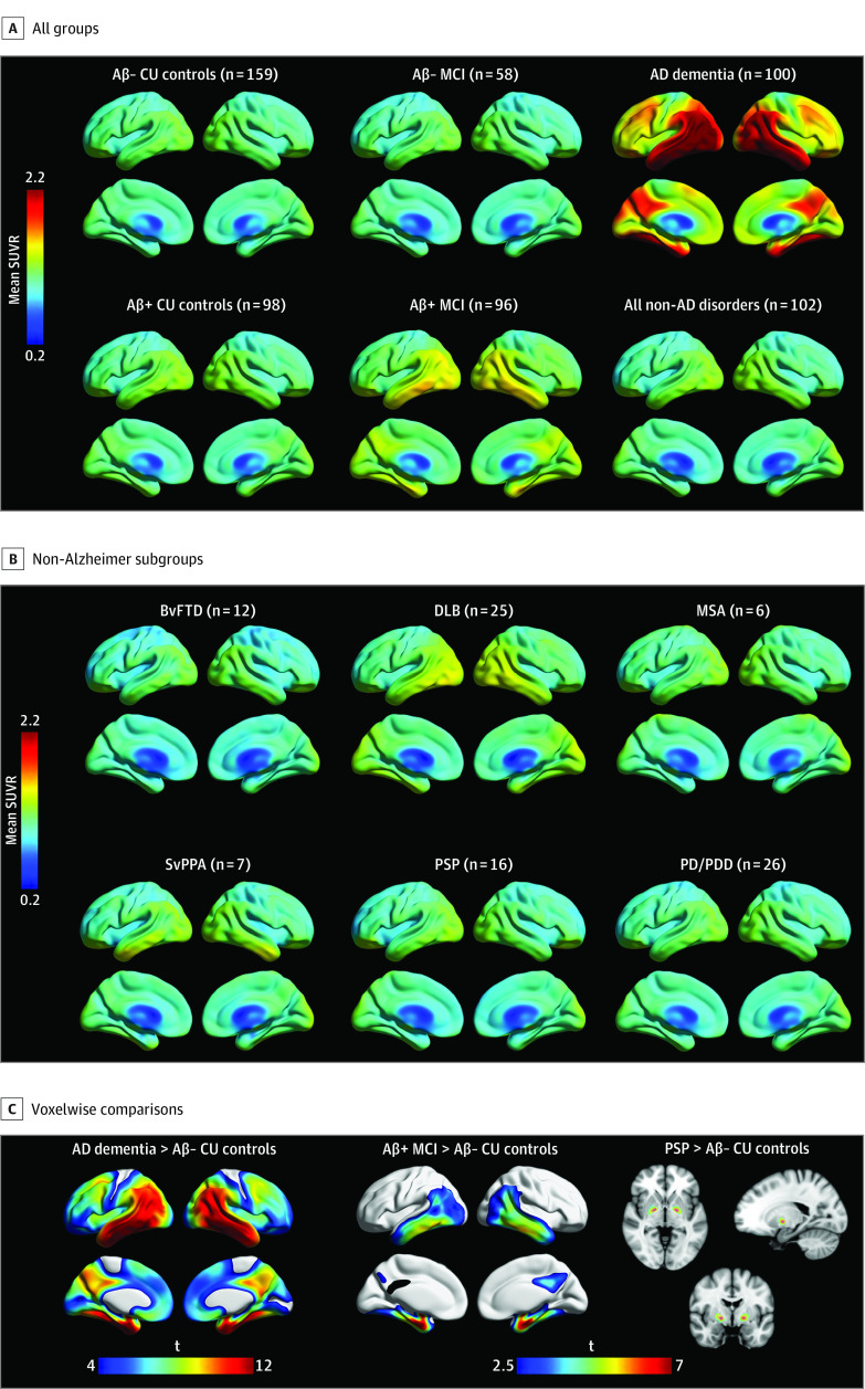
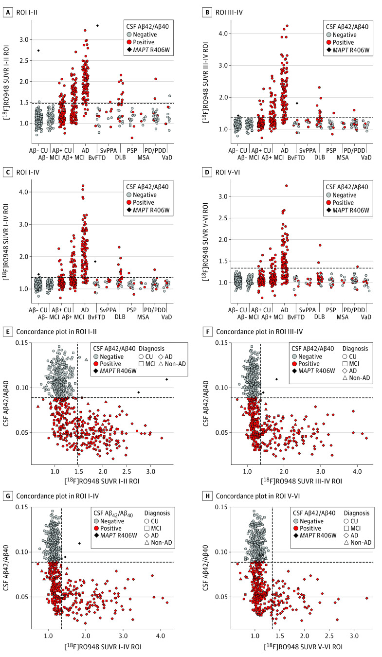
Main outcomes and measures: Standard uptake value ratios (SUVRs) in 4 predefined regions of interest (ROIs) reflecting Braak staging scheme for tau pathology and encompass I-II (entorhinal cortex), III-IV (inferior/middle temporal, fusiform gyrus, parahippocampal cortex, and amygdala), I-IV, and V-VI (widespread neocortical areas), area under the receiver operating characteristic curve (AUC) values, and subtraction images between [18F]RO948 and [18F]flortaucipir.

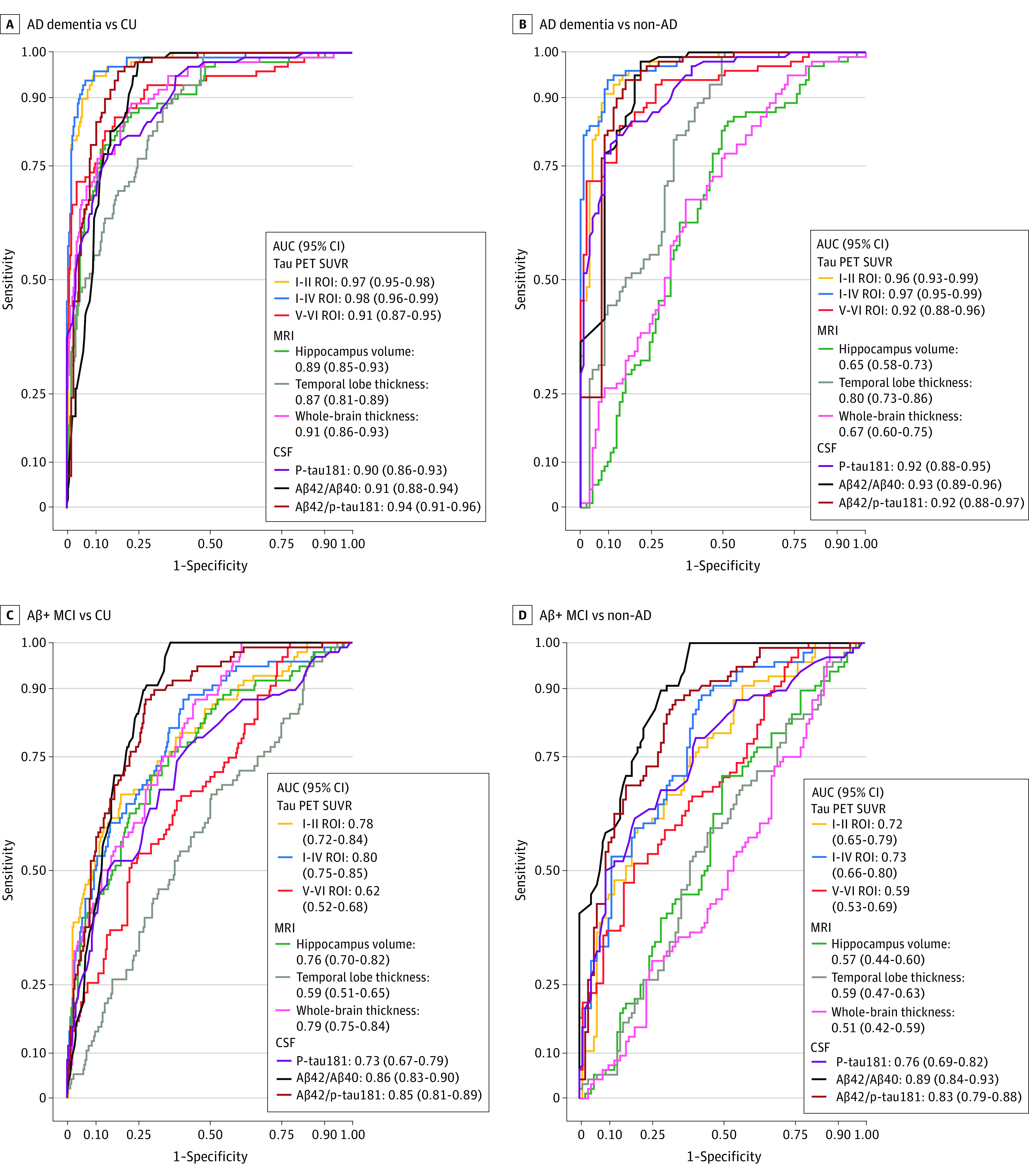
Results: Diagnostic groups among the 613 participants included cognitively unimpaired (mean [SD] age, 65.8 [12.1] years; 117 men [46%]), mild cognitive impairment (age, 70.8 [8.3] years; 82 men [53%]), AD dementia (age, 73.5 [6.7] years; 57 men [57%]), and non-AD disorders (age, 70.5 [8.6] years; 41 men [40%]). Retention of [18F]RO948 was higher in AD dementia compared with all other diagnostic groups. [18F]RO948 could distinguish patients with AD dementia from individuals without cognitive impairment and those with non-AD disorders, and the highest AUC was obtained using the I-IV ROI (AUC = 0.98; 95% CI, 0.96-0.99 for AD vs no cognitive impairment and AUC = 0.97; 95% CI, 0.95-0.99 for AD vs non-AD disorders), which outperformed MRI (highest AUC = 0.91 for AD vs no cognitive impairment using whole-brain thickness, and AUC = 0.80 for AD vs non-AD disorders using temporal lobe thickness) and cerebrospinal fluid measures (highest AUC = 0.94 for AD vs no cognitive impairment using Aβ42/p-tau181, and AUC = 0.93 for AD vs non-AD disorders using Aβ42/Aβ40). Generally, tau PET positivity using [18F]RO948 was observed only in Aβ-positive cases or in MAPT R406W mutation carriers. Retention of [18F]RO948 was not pronounced in patients with svPPA, and head-to-head comparison revealed lower temporal lobe uptake than with [18F]flortaucipir.

Conclusions and relevance: In this study, elevated [18F]RO948 SUVRs were most often seen among Aβ-positive cases, which suggests that [18F]RO948 has high specificity for AD-type tau and highlights its potential as a diagnostic marker in the differential diagnosis of AD.


Conflict of interest statement

Conflict of Interest Disclosures: Dr Santillo reported receiving grants from the Swedish Society for Medical Research and the Bente Rexhed Gerstedts Foundation for Medical Research during the conduct of the study. Dr Klein reported receiving personal fees from F. Hoffmann-La Roche Ltd during the conduct of the study. Dr Hansson reported receiving grants from Roche during the conduct of the study as well as grants from Roche, nonfinancial support from GE Healthcare, and grants from Biogen outside the submitted work. No other disclosures were reported.

Figures

Figure 1.
Figure 1.. Mean RO948 F 18 ([18F]RO948) Standardized Uptake Value Ratio (SUVR) Images Across Diagnostic Groups
A, Cognitively unimpaired (CU) control individuals and patients with mild cognitive impairment (MCI) and Alzheimer disease (AD) dementia. AD dementia was greater than amyloid β (Aβ)-negative CU controls and Aβ-positive MCI. B, Non-AD subgroups compared with Aβ-negative CU controls. Aβ vs APOE ε4 status (% positive) by non-AD subgroup was as follows: behavioral variant of frontotemporal dementia (BvFTD) (50% vs 33%), dementia with Lewy bodies (DLB) (60% vs 52%), multiple system atrophy (MSA) (33% vs 33%), semantic variant primary progressive aphasia (svPPA) (71% vs 17%), progressive supranuclear palsy (PSP) (38% vs 31%), and Parkinson disease/Parkinson disease dementia (PD/PDD) (23% vs 27%). C, Voxelwise group differences in [18F]RO948 SUVRs. AD dementia, Aβ-positive MCI, and PSP are compared with Aβ-negative CU controls. A cluster threshold of 100 voxels was applied with no correction for multiple comparisons (P < .001). t indicates t value.
Figure 2.
Figure 2.. Mean RO948 F 18 ([18F]RO948) Standardized Uptake Value Ratios (SUVRs) Across Diagnostic Groups Within Tau-Imaging Regions of Interest (ROIs)
ROI groupings were I-II (A), III-IV (B), I-IV (C), and V-VI (D). Concordance plots are shown between the [18F]RO948 SUVR and cerebrospinal fluid (CSF) amyloid β42 and β40 (Aβ42/Aβ40) for ROIs I-II (E), III-IV (F), I-IV (G), and V-VI (H). The horizontal dashed lines indicate the cutoffs for tau positivity across the ROIs, defined using the mean plus 2.5 SDs in Aβ-negative young controls (I-II ROI>1.48; III-IV and I-IV ROIs >1.36; V-VI ROI>1.35). The vertical dashed line indicates the cutoff for Aβ-positivity (CSF Aβ42/Aβ40<0.089, as established in an independent population by the neurochemistry laboratory at the Sahlgrenska University Hospital, Mölndal, Sweden). Abbreviation expansions appear in caption to Figure 1. VaD indicates vascular dementia.
Figure 3.
Figure 3.. Plots From Receiver Operating Characteristic Analyses for RO948 F 18 ([18F]RO948) Tau Positron Emission Tomographic (PET) Tracers, Magnetic Resonance Imaging (MRI), and Cerebrospinal Fluid (CSF) Measures for Distinguishing Alzheimer Disease (AD) Dementia and Amyloid β (Aβ)-Positive Mild Cognitive Impairment (MCI) From Cognitively Unimpaired Controls and Non-AD Neurodegenerative Disorders
Receiver operating characteristic curves are shown for the following groups: AD dementia vs cognitively unimpaired (CU) controls (A) and non-AD disorders (B), and Aβ-positive MCI vs CU controls (C) and non-AD disorders (D). The III-IV region of interest, omitted owing to its similar performance to I-IV, had the following area under the curve (AUC) values: AD dementia vs cognitively unimpaired controls, AUC = 0.97 (95% CI, 0.95-0.99); AD dementia vs non-AD: AUC = 0.96 (95% CI, 0.94-0.98); Aβ-positive MCI vs cognitively unimpaired controls: AUC = 0.77 (95% CI, 0.72-0.83); and Aβ-positive MCI vs non-AD: AUC = 0.71 (95% CI, 0.65-0.78). ROI indicates region of interest; SUVR, standardized uptake value ratio.

References

    1. Blennow K, de Leon MJ, Zetterberg H. Alzheimer’s disease. Lancet. 2006;368(9533):387-403. doi:10.1016/S0140-6736(06)69113-7 - DOI - PubMed
    1. Jellinger KA, Bancher C. Neuropathology of Alzheimer’s disease: a critical update. J Neural Transm Suppl. 1998;54:77-95. doi:10.1007/978-3-7091-7508-8_8 - DOI - PubMed
    1. Iqbal K, Liu F, Gong CX, Grundke-Iqbal I. Tau in Alzheimer disease and related tauopathies. Curr Alzheimer Res. 2010;7(8):656-664. doi:10.2174/156720510793611592 - DOI - PMC - PubMed
    1. Lee VM, Goedert M, Trojanowski JQ. Neurodegenerative tauopathies. Annu Rev Neurosci. 2001;24:1121-1159. doi:10.1146/annurev.neuro.24.1.1121 - DOI - PubMed
    1. Goedert M. Tau filaments in neurodegenerative diseases. FEBS Lett. 2018;592(14):2383-2391. doi:10.1002/1873-3468.13108 - DOI - PubMed

Publication types

MeSH terms