Review
. 2020 Sep 1;58(3):500-510.
doi: 10.1093/ejcts/ezaa093.

Current challenges in three-dimensional bioprinting heart tissues for cardiac surgery

Affiliations
Review

Current challenges in three-dimensional bioprinting heart tissues for cardiac surgery

Christopher D Roche et al. Eur J Cardiothorac Surg. .

Abstract

Previous attempts in cardiac bioengineering have failed to provide tissues for cardiac regeneration. Recent advances in 3-dimensional bioprinting technology using prevascularized myocardial microtissues as 'bioink' have provided a promising way forward. This review guides the reader to understand why myocardial tissue engineering is difficult to achieve and how revascularization and contractile function could be restored in 3-dimensional bioprinted heart tissue using patient-derived stem cells.

Keywords: Bioprinting; Cardiac tissues; Regenerative medicine; Revascularization; Stem cells; Transplantation.


Figures

Figure 1:
Figure 1:
Extrusion-based 3D bioprinting and cell viability. (A) Four-nozzle extrusion 3D bioprinting system using pneumatic force to extrude 4 different bioinks. In this example, bioinks are extruded directly into a crosslinker solution, which acts on the bioink to create bonds within the bioprinted structure to retain its shape. (B) Downward arrows with greater size indicate greater velocity centrally in the bioink and lower velocity at the periphery, which is in contact with the chamber wall. Upward arrows show resulting shear stress on cells in bioink during 3D bioprinting. Greater shear at the periphery results in stressed cells (orange) and some dead cells (red). Shear stress can be reduced by slowing down the velocity of extrusion, which is readily controlled in 3D bioprinting (reproduced with permission from Blaeser et al. [18]). 3D: 3-dimensional.
Figure 2:
Figure 2:
Three-dimensional cardiac microtissue generation and physiology. (A) Standard 2-dimensional cell culture of human-induced pluripotent stem cell-derived cardiomyocytes. (B) Single (∼200 μm diameter) cardiac spheroid microtissue—a 3-dimensional aggregate of cardiac cells with ability to control spheroid size and cell number and optimize cell–cell interactions, 3-dimensional mechanical signals and extracellular support. (C) Two methods for generating cardiac spheroids: U-shaped non-adhesive wells (top panel) and hanging drop cultures (bottom panel). (D) Two types of field pacing chambers for providing electrical stimulation to spheroids for the optimal maturation of cardiomyocyte phenotype: a perfusion chamber (above) and culture dish (below) with electrodes to allow for field potential stimulation across cardiac spheroid culture area (modified with permission from Zuppinger [1]).
Figure 3:
Figure 3:
Vascularization in tissue-engineered cardiac patches. (A) CD31 expression (green) in a cardiac patch generated by a fibrinogen moulding method and engrafted in pigs (modified with permission from Gao et al. [4]). (B) CD31 expression (green) in a cardiac patch generated by a net moulding method and engrafted in rats (modified with permission from Yang et al. [19]). (C) CD31 expression (red) in a fibrin-based cardiac patch generated by a spheroid fusion method and engrafted in mice (modified with permission from Mattapally et al. [6]). (D) CD31 expression (green) in a cardiac patch generated by a 3-dimensional bioprinting method (modified with permission from Noor et al. [5]). (E) CD31 expression (red) in a cardiac patch generated by a 3-dimensional bioprinting method (modified with permission from Zhang et al. [10]). (F) vWF (red) and lamin A/C (green) expressions in a cardiac patch generated by a 3-dimensional bioprinting method and engrafted in mice with the evidence of host-patch anastomosis (modified with permission from Maiullari et al. [9]). Scale bar appearances are due to source data. vWF: von Willebrand factor.
Figure 4:
Figure 4:
Electrical stimulation of cell culture leads to greater contractility in engineered cardiac tissue. Beating macrotissues were generated by seeding induced pluripotent stem cell-derived cardiomyocytes and human fibroblasts in a 3-dimensional porous scaffold and culturing for 14 days as either control (no electrical stimulation) or ‘CardioSlice’ (electrical stimulation applied whilst culturing). (A) Human cardiac macrotissues after 7 or 14 days of culture, either without (control) or with (CardioSlice) electrical stimulation (scale bars 2.5 mm) (see also Videos 1 and 2). (B) Contraction amplitude of control versus CardioSlice bioengineered cardiac tissues. Fractional contraction area (compared to area of the tissue at rest) is represented over time, and control tissue remains close to 1.0 when contracting whereas CardioSlice patches contract to 0.85 (85% of size of tissue at rest). (C) The percentage of FAC for control (unstimulated) versus electrically stimulated CardioSlice cardiac macrotissues. (D) MCR for control compared to electrically stimulated CardioSlice macrotissues when paced. At 14 days, CardioSlice macrotissues were able to be paced at over 4 Hz, approximately double the MCR of control tissues. (E) Beating cardiac macrotissue velocity maps after 14 days of culture. Blue colours and shorter white mini arrows represent lower velocities. Higher velocities (redder areas and longer mini arrows) were observed in CardioSlice macrotissues versus controls (scale bar 2.5 mm). (F) Alignment analysis comparing direction of the electrical field vector and subsequent beating direction of bioengineered cardiac tissues. The order parameter cos2θ was used: random distribution gives values close to 0 whereas parallel alignment gives values close to 1 (modified with permission from Valls-Margarit et al. [21]). FAC: fractional area change; MCR: maximum capture rate.
Figure 5:
Figure 5:
Embryonic stem cell-derived cardiac patches for patients with heart failure undergoing coronary artery bypass grafting (CABG). (A–C) A fibrin-based patch infused with allogenic cardiac progenitor cells derived from embryonic stem cells was applied to the epicardial surface of human cardiac failure patients undergoing CABG and shown to be safe in a phase I clinical safety trial (modified with permission from Menasché et al. [33]).
Figure 6:
Figure 6:
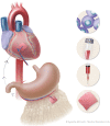
The human body as a natural bioreactor for bioprinted cardiac patch vascularization. Patient-specific induced pluripotent stem cell-derived bioinks obtained by reprogramming cells taken from patient blood could be used to 3-dimensional bioprint cardiac tissue patches. This tissue could be matured on the patient’s omentum, anastomosed to the gastroepiploic artery and subsequently rotated onto the epicardial surface to regenerate the myocardium—a waiting list-free and surgically feasible alternative to donor heart allotransplantation.
None

References

    1. Zuppinger C.Measurement of contractility and calcium release in cardiac spheroids. Methods Mol Biol 2019;1929:41–52. - PubMed
    1. Beyersdorf F.Three-dimensional bioprinting: new horizon for cardiac surgery. Eur J Cardiothorac Surg 2014;46:339–41. - PubMed
    1. Puluca N, Lee S, Doppler S, Münsterer A, Dreßen M, Krane M. et al.Bioprinting approaches to engineering vascularized 3D cardiac tissues. Curr Cardiol Rep 2019;21:90. - PMC - PubMed
    1. Gao L, Gregorich ZR, Zhu W, Mattapally S, Oduk Y, Lou X. et al.Large cardiac muscle patches engineered from human induced-pluripotent stem cell-derived cardiac cells improve recovery from myocardial infarction in swine. Circulation 2018;137:1712–30. - PMC - PubMed
    1. Noor N, Shapira A, Edri R, Gal I, Wertheim L, Dvir T.. 3D printing of personalized thick and perfusable cardiac patches and hearts. Adv Sci 2019;6:1900344. - PMC - PubMed

Publication types