Review
. 2021 Mar;10(2):e382.
doi: 10.1002/wdev.382. Epub 2020 May 11.

Long-distance regressive signaling in neural development and disease

Affiliations
Review

Long-distance regressive signaling in neural development and disease

Amrita Pathak et al. Wiley Interdiscip Rev Dev Biol. 2021 Mar.

Abstract

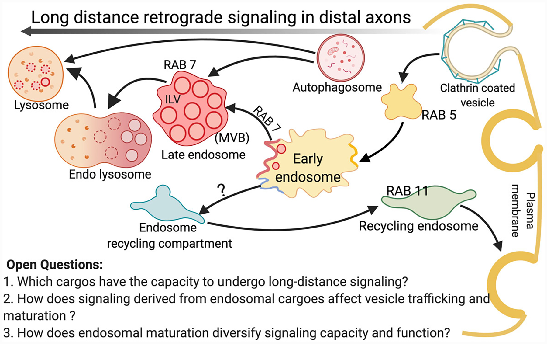
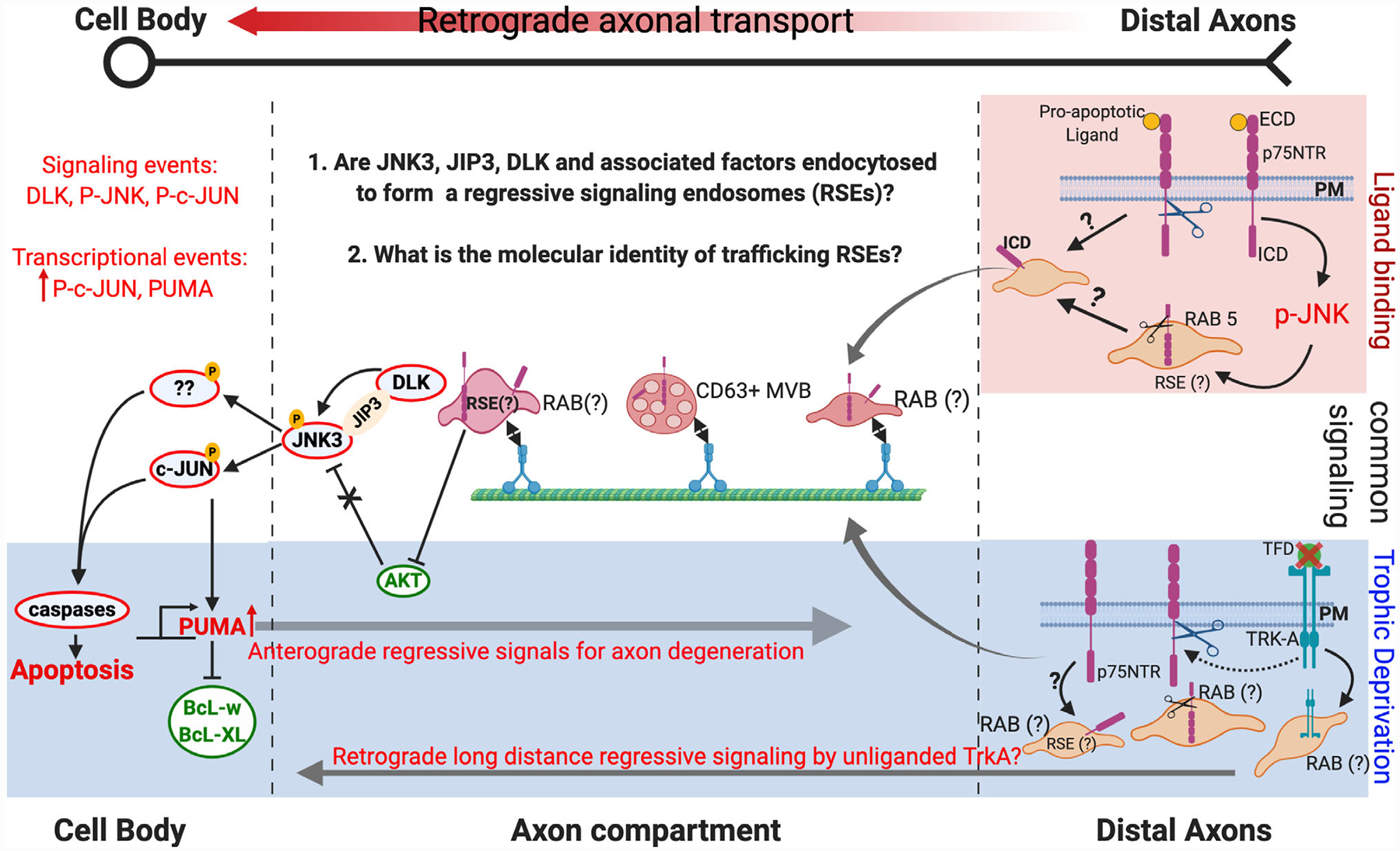
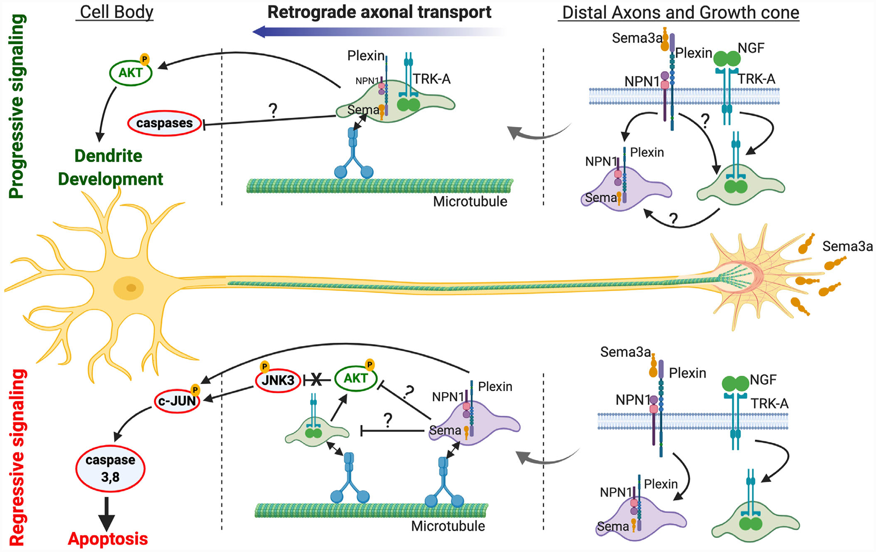
Nervous system development proceeds via well-orchestrated processes involving a balance between progressive and regressive events including stabilization or elimination of axons, synapses, and even entire neurons. These progressive and regressive events are driven by functionally antagonistic signaling pathways with the dominant pathway eventually determining whether a neural element is retained or removed. Many of these developmental sculpting events are triggered by final target innervation necessitating a long-distance mode of communication. While long-distance progressive signaling has been well characterized, particularly for neurotrophic factors, there remains relatively little known about how regressive events are triggered from a distance. Here we discuss the emergent phenomenon of long-distance regressive signaling pathways. In particular, we will cover (a) progressive and regressive cues known to be employed after target innervation, (b) the mechanisms of long-distance signaling from an endosomal platform, (c) recent evidence that long-distance regressive cues emanate from platforms like death receptors or repulsive axon guidance receptors, and (d) evidence that these pathways are exploited in pathological scenarios. This article is categorized under: Nervous System Development > Vertebrates: General Principles Signaling Pathways > Global Signaling Mechanisms Establishment of Spatial and Temporal Patterns > Cytoplasmic Localization.

Keywords: axon transport; degeneration; neurotrophin; p75NTR; signaling.


Conflict of interest statement

CONFLICT OF INTEREST

The authors have declared no conflicts of interest for this article.

Figures

FIGURE 1
FIGURE 1
Vesicles involved in retrograde axonal transport and signaling. After ligand binding receptors are typically internalized at the plasma membrane of distal axons. RAB5 or early endosome Antigen1 (EEA1) is a marker for early endosomes where the cargo is sorted into different endocytic compartments, while RAB7 is thought to control the maturation of late endosomes, their retrograde trafficking, and fusion to lysosomes. RAB5 or RAB7 labeled vesicles, lysosomes, and autophagosomes have all been observed to undergo retrograde transport. The intralumenal vesicles (ILVs) accumulate in the lumen of endosomes, which consequently change their composition and morphology to convert into a multivesicular body (MVB), which can also be retrogradely transported
FIGURE 2
FIGURE 2
Comparison of regressive signaling under pro-apoptotic ligand binding and trophic factor deprivation (TFD). Top shade (red) depicts signaling after pro-apoptotic ligand binding to p75NTR, white area depicts common signaling while the bottom shade (blue) indicates signals identified under trophic factor deprivation (TFD) due to NGF withdrawal. Scissors indicate cleavage by γ-secretase to release the intracellular domain (ICD) of the receptor. Red outlined circles indicate regressive signaling components and Green outlined circles represent progressive signaling components. PM, plasma membrane; ECD, extracellular domain; ICD, intracellular domain; RSE, regressive signaling endosome. The sequence of events for p75NTR endocytosis and cleavage by γ-secretase in distal axons is not known. p75NTR does not function in a regressive manner in all cellular contexts (e.g., sensory neurons) and unliganded TRK-A has been shown to signal (also see section 4 B.2 on dependence receptors). Whether unliganded TRK-A is a component of RSE or contributes to p75NTR mediated long-distance regressive signaling remains to be tested
FIGURE 3
FIGURE 3
Long-distance signaling by Semaphorin 3a. Semaphorin 3A (Sema3A) exerts its biological actions through receptors, neuropilin-1 (Npn1) and plexin family members. For dendrite development, Sema3a are shown to induce PlexA4 colocalization and interaction with TRK-A receptor in growth cones and along axons (N. Yamashita et al., 2016). For regressive signaling, Sema3a signals retrograde apoptosis in developing sympathetic neurons via PlexinA3-NPN-1 which requires caspase 3 and 8 but does not depend on the availability of NGF on distal axons (Wehner et al., 2016). The untested hypothetical segments of retrograde signaling by Sema3a are indicated by question marks. Sema3a is depicted as sema due to space limitation

References

    1. Ascano M, Bodmer D, & Kuruvilla R (2012). Endocytic trafficking of neurotrophins in neural development. Trends in Cell Biology, 22(5), 266–273. 10.1016/j.tcb.2012.02.005 - DOI - PMC - PubMed
    1. Ayloo S, Lazarus JE, Dodda A, Tokito M, Ostap EM, & Holzbaur ELF (2014). Dynactin functions as both a dynamic tether and brake during dynein-driven motility. Nature Communications, 5, 4807 10.1038/ncomms5807 - DOI - PMC - PubMed
    1. Baleriola J, Walker CA, Jean YY, Crary JF, Troy CM, Nagy PL, & Hengst U (2014). Axonally synthesized ATF4 transmits a neurodegenerative signal across brain regions. Cell, 158(5), 1159–1172. 10.1016/j.cell.2014.07.001 - DOI - PMC - PubMed
    1. Ballard C, Mobley W, Hardy J, Williams G, & Corbett A (2016). Dementia in Down’s syndrome. Lancet Neurology, 15(6), 622–636. 10.1016/S1474-4422(16)00063-6 - DOI - PubMed
    1. Barford K, Deppmann C, & Winckler B (2017). The neurotrophin receptor signaling endosome: Where trafficking meets signaling. Developmental Neurobiology, 77(4), 405–418. 10.1002/dneu.22427 - DOI - PMC - PubMed

FURTHER READING

    1. Aravamudhan P, Raghunathan K, Konopka-Anstadt J, Pathak A, Sutherland DM, Carter BD, & Dermody TS (2020). Reovirus uses macropinocytosis-mediated entry and fast axonal transport to infect neurons. PLOS Pathogens, 16(2), e1008380 10.1371/journal.ppat.1008380 - DOI - PMC - PubMed
    1. Fantuzzo JA, Hart RP, Zahn JD, & Pang ZP (2019). Compartmentalized devices as tools for investigation of human brain network dynamics. Developmental Dynamics, 248, 65–77. https://doi.org/10.1002/dvdy.24665 Retrieved from https://doi.org/10.1002/dvdy.24665https://anatomypubs.onlinelibrary.wiley.com/doi/pdf/10.1002/dvdy.24665 Retrieved from https://anatomypubs.onlinelibrary.wiley.com/doi/pdf/10.1002/dvdy.24665 - DOI - DOI - PMC - PubMed
    1. Langemeyer L, Fröhlich F, & Ungermann C (2018). Rab GTPase function in endosome and lysosome biogenesis. Trends in Cell Biology, 28(11), 957–970. https://doi.org/10.1016/j.tcb.2018.06.007 Retrieved from https://doi.org/10.1016/j.tcb.2018.06.007https://www.sciencedirect.com/science/article/pii/S0962892418301089?via%... Retrieved from https://www.sciencedirect.com/science/article/pii/S0962892418301089?via%... - DOI - PubMed
    1. Lawrence RE, & Zoncu R (2019). The lysosome as a cellular centre for signalling, metabolism and quality control. Nature Cell Biology, 21, 133–142. https://doi.org/10.1038/s41556-018-0244-7 Retrieved from https://doi.org/10.1038/s41556-018-0244-7https://www.nature.com/articles/s41556-018-0244-7 Retrieved from https://www.nature.com/articles/s41556-018-0244-7 - DOI - PubMed
    1. Martin N, Gonzalo R, Stephanie M, Anne S, Raimund S, Kyoohyun K, … Jochen G (2018). Axonal transport, phase-separated compartments, and neuron mechanics—A new approach to investigate neurodegenerative diseases. Frontiers in Cellular Neuroscience, 12, 358 https://doi.org/10.3389/fncel.2018.00358 Retrieved from https://doi.org/10.3389/fncel.2018.00358https://www.frontiersin.org/articles/10.3389/fncel.2018.00358/full Retrieved from https://www.frontiersin.org/articles/10.3389/fncel.2018.00358/full - DOI - DOI - PMC - PubMed

Publication types

Substances