Review
. 2021;17(2):173-181.
doi: 10.2174/1573403X16666200511090151.

Conduction Disorders: The Value of Surface ECG

Affiliations
Review

Conduction Disorders: The Value of Surface ECG

Rose M F L da Silva et al. Curr Cardiol Rev. 2021.

Abstract

Purpose of review: The purpose of the current mini-review is to describe the importance of surface ECG for the diagnosis of conduction disorder.

Methods: The MEDLINE/PubMed database was used, with the keywords "ECG" and "conduction disorders"; over the past 10 years. Other documents were included because of their relevance.

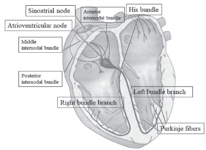
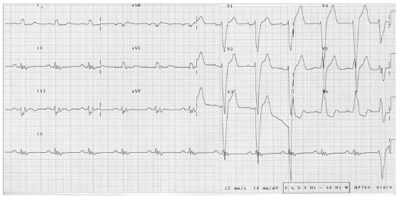
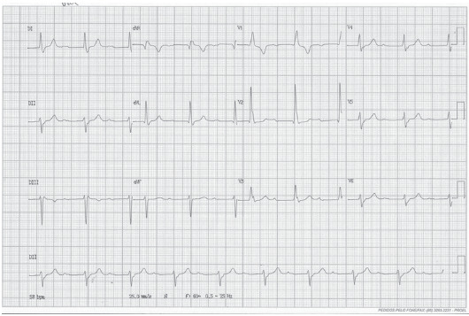
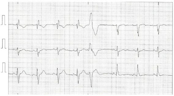
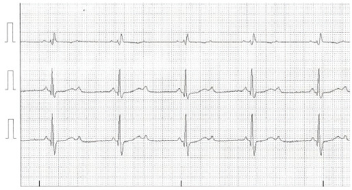
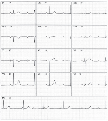
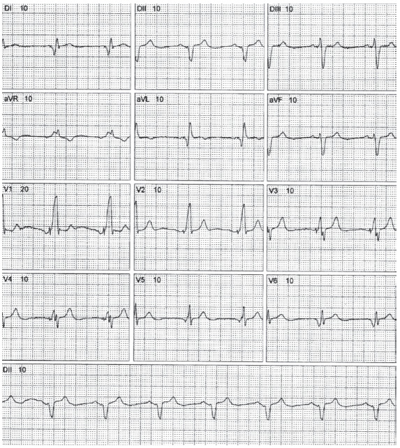
Main findings: Data on the anatomy and function of the cardiac electrical system have been described. Conduction disorders including sinus node dysfunction, atrioventricular blocks, intraventricular conduction disorders are exposed as to their epidemiology, etiology, presentation, anatomical site of impaired conduction of the electrical stimulus. The importance of ECG in patients with a cardiac implantable electronic device was also discussed, in addition to future perspectives.

Conclusion: Surface ECG allows the diagnosis of atrioventricular and intraventricular conduction disorder and its anatomical block site most of the time, without the need for invasive tests such as electrophysiological study. Dysfunctions of cardiac implantable electronic devices can be diagnosed by ECG, as well as the prediction of response to cardiac resynchronization therapy.

Keywords: Cardiac conduction system diseases; bundle-branch block; cardiac resynchronization therapy.; electrocardiography; heart conduction system; sinus node dysfunction.


Figures

Fig. (1)
Fig. (1)
Main structures of cardiac conduction system. (A higher resolution / colour version of this figure is available in the electronic copy of the article).
Fig. (2)
Fig. (2)
Electrocardiographic tracing of leads V1, V5 and D3 showing ventricular pause of 4.2 seconds in a 75-year-old patient with syncope. (A higher resolution / colour version of this figure is available in the electronic copy of the article).
Fig. (3)
Fig. (3)
12-lead electrocardiographic tracing demonstrating sinus rhythm with complete left bundle branch block. There is also QRS fragmentation in the inferior leads. This fragmentation represents conduction delay due to myocardial scarring and is a marker of cardiovascular disease risk [22]. (A higher resolution / colour version of this figure is available in the electronic copy of the article).
Fig. (4)
Fig. (4)
12-lead electrocardiographic tracing demonstrating sinus rhythm with bifascicular block (right bundle branch block and left anterior fascicular block) with prolonged PR. (A higher resolution / colour version of this figure is available in the electronic copy of the article).
Fig. (5)
Fig. (5)
Electrocardiographic tracing of modified V1, V5, and D3 leads demonstrating sinus rhythm with alternating bundle branch block, right bundle branch block at the beginning of the ECG, followed by ventricular extrasystole and left bundle branch block. (A higher resolution / colour version of this figure is available in the electronic copy of the article).
Fig. (6)
Fig. (6)
Electrocardiographic tracing of modified V1 and V5 leads and D3, demonstrating 2:1 AV block with right bundle branch block. Thus, the anatomical site of the block is infra-His. (A higher resolution / colour version of this figure is available in the electronic copy of the article).
Fig. (7)
Fig. (7)
12-lead electrocardiographic tracing demonstrating sinus atrial rhythm, total AV block with narrow QRS complex, with two sinus capture beats (4th and 5th QRS complexes of the D2 lead tracing at the bottom). (A higher resolution / colour version of this figure is available in the electronic copy of the article).
Fig. (8)
Fig. (8)
12-lead ECG of 72-year-old male patients with response to cardiac resynchronization therapy. (A higher resolution / colour version of this figure is available in the electronic copy of the article).

References

    1. Fye W.B. A history of the origin, evolution, and impact of electrocardiography. Am. J. Cardiol. 1994;73(13):937–949. doi: 10.1016/0002-9149(94)90135-X. - DOI - PubMed
    1. AlGhatrif M., Lindsay J. A brief review: History to understand fundamentals of electrocardiography. J. Community Hosp. Intern. Med. Perspect. 2012;2(1) doi: 10.3402/jchimp.v2i1.14383. - DOI - PMC - PubMed
    1. de Luna A.B. Willem Einthoven and the ECG. Eur. Heart J. 2019;40(41):3381–3383. doi: 10.1093/eurheartj/ehz721. - DOI - PubMed
    1. Haberman Z.C., Jahn R.T., Bose R., et al. Wireless smartphone ECG enables large-scale screening in diverse populations. J. Cardiovasc. Electrophysiol. 2015;26(5):520–526. doi: 10.1111/jce.12634. - DOI - PubMed
    1. Costantini O. Basic principles of cardiac electrophysiology. Med. Clin. North Am. 2019;103(5):767–774. doi: 10.1016/j.mcna.2019.04.002. - DOI - PubMed

MeSH terms