. 2020 May 11;16(5):e1008576.
doi: 10.1371/journal.ppat.1008576. eCollection 2020 May.

Yersinia pseudotuberculosis YopH targets SKAP2-dependent and independent signaling pathways to block neutrophil antimicrobial mechanisms during infection

Affiliations

Yersinia pseudotuberculosis YopH targets SKAP2-dependent and independent signaling pathways to block neutrophil antimicrobial mechanisms during infection

Lamyaa Shaban et al. PLoS Pathog. .

Abstract

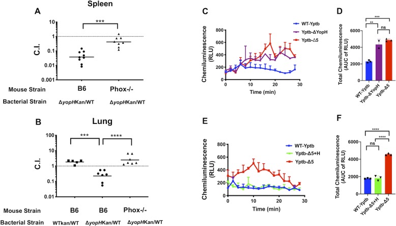
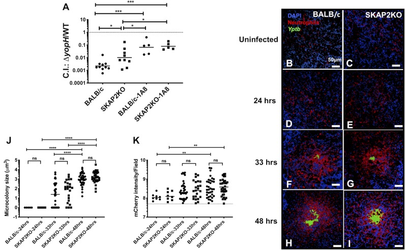
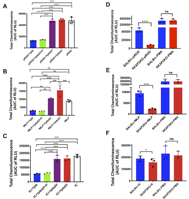
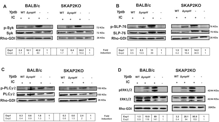
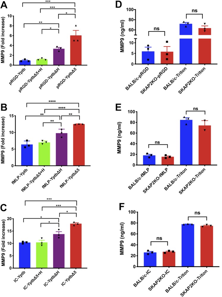
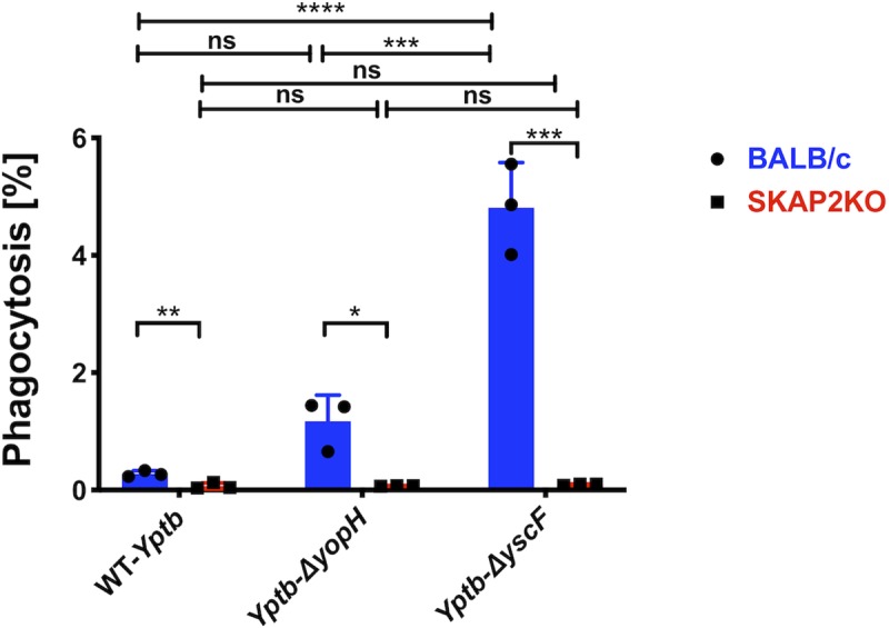
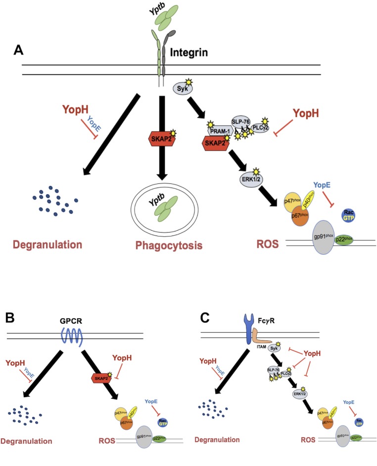
Yersinia suppress neutrophil responses by using a type 3 secretion system (T3SS) to inject 6-7 Yersinia effector proteins (Yops) effectors into their cytoplasm. YopH is a tyrosine phosphatase that causes dephosphorylation of the adaptor protein SKAP2, among other targets in neutrophils. SKAP2 functions in reactive oxygen species (ROS) production, phagocytosis, and integrin-mediated migration by neutrophils. Here we identify essential neutrophil functions targeted by YopH, and investigate how the interaction between YopH and SKAP2 influence Yersinia pseudotuberculosis (Yptb) survival in tissues. The growth defect of a ΔyopH mutant was restored in mice defective in the NADPH oxidase complex, demonstrating that YopH is critical for protecting Yptb from ROS during infection. The growth of a ΔyopH mutant was partially restored in Skap2-deficient (Skap2KO) mice compared to wild-type (WT) mice, while induction of neutropenia further enhanced the growth of the ΔyopH mutant in both WT and Skap2KO mice. YopH inhibited both ROS production and degranulation triggered via integrin receptor, G-protein coupled receptor (GPCR), and Fcγ receptor (FcγR) stimulation. SKAP2 was required for integrin receptor and GPCR-mediated ROS production, but dispensable for degranulation under all conditions tested. YopH blocked SKAP2-independent FcγR-stimulated phosphorylation of the proximal signaling proteins Syk, SLP-76, and PLCγ2, and the more distal signaling protein ERK1/2, while only ERK1/2 phosphorylation was dependent on SKAP2 following integrin receptor activation. These findings reveal that YopH prevents activation of both SKAP2-dependent and -independent neutrophilic defenses, uncouple integrin- and GPCR-dependent ROS production from FcγR responses based on their SKAP2 dependency, and show that SKAP2 is not required for degranulation.


Conflict of interest statement

The authors have declared that no competing interests exist.

Figures

Fig 1
Fig 1. The growth of a ΔyopH mutant is restored in absence of ROS in vivo and YopH is necessary and sufficient to block extracellular ROS produced by BM neutrophils in vitro.
(A-B) Female C57BL/6J or C57BL/6J gp91phox-/- mice were inoculated with a 1:1 mixture of IP2666 WT-Yptb and ΔyopH-KanR (A) intravenously with 1x10³ CFU or (B) intratracheally with 5x10³ CFU. Spleens and lungs were collected, weighed, homogenized, and plated for CFU two days post- infection. The competitive indices (C.I.) were determined by patching 100 colonies on L-Irgasan and L-Irgasan+Kanamycin plates. Each symbol represents an individual mouse; horizontal bars represent the geometric mean. (C-F) BM neutrophils were infected at a MOI of 20:1 with (C-D) WT-Yptb, ΔyopH, or Δ5 (a mutant lacking 5 effector Yops, H, E, O, M, J), (E-F) WT-Yptb+pBAD, Δ5+YopH (a strain expressing only YopH at native levels from an arabinose inducible plasmid),or Δ5+pBAD, and monitored for extracellular ROS production using isoluminol chemiluminescence assay. (D and F) Total chemiluminescence was determined by computing the area under the curve (AUC) using GraphPad Prism version 7. Mean ±SD of a representative experiment from 2–3 independent experiments done in 2–3 replicates is presented. Statistical significance was calculated using (A) the Mann-Whitney t-test, (B) one-way ANOVA followed by Tukey’s Post-test after log10 transformation of the data, or (D, F) one-way ANOVA followed by Tukey’s Post-test for area under the curve (AUC).
Fig 2
Fig 2. Skap2KO neutrophils cluster to Yptb microcolonies and the growth of a ΔyopH mutant is partially restored in competition with WT-Yptb in Skap2KO mice.
(A) WT-BALB/c and Skap2KO mice were injected with 1A8 or an isotype control antibody and 16 hrs later I.V. infected with an equal mixture of 1x10³ CFU of IP2666 WT-Yptb and ΔyopH-KanR. Spleens were collected three days post-infection, weighed, homogenized, and plated for CFU on selective and non-selective agar. The number of bacteria recovered from selective and non-selective plates was used to determine the C.I. Each dot represents a mouse; horizontal bars represent the geometric mean. Significance was calculated using 2-way ANOVA followed by Tukey’s Post-test of log10 transformed values, with only significant values shown. The results are a composite of three experiments. (B-I) BALB/c and Skap2KO mice were (B-C) uninfected or (D-I) I.V. infected with 103 CFU of IP2666 WT-Yptb-GFP (green), and sacrificed at (D-E) 24, (F-G) 33, or (H-I) 48 hours post-infection. Frozen sections of spleens were stained using an anti-Ly6G antibody (neutrophils-red) and DAPI (nuclei-blue) and visualized by fluorescence confocal microscopy at 40X magnification. (J-K) Using Volocity software, the size of the (J) bacterial microcolony was determined by generating the summed volume of the individual GFP signal and the (K) number of neutrophils recruited was determined by detecting the signal intensity of the mCherry channel. Each dot represents a microcolony or an area of clustered neutrophils. The horizontal solid bars represent the median, and (K) the dotted line represents the median neutrophil signal intensity in uninfected controls of BALB/c and Skap2KO mice. Scale bars: 50 μm. Statistical significance was determined (J-K) using Mann-Whitney for comparing BALB/c and Skap2KO at each time point and Kruskal-Wallis for comparisons between the three time points for each mouse genotype. The results are a composite of 2–3 mice from two independent experiments.
Fig 3
Fig 3. YopH is necessary to block ROS from all three receptors while Skap2KO neutrophils fail to produce ROS following integrin receptor and GPCR but not FcγR stimulation.
Respiratory burst of (A-C) WT BM neutrophils that were either uninfected or infected with IP2666 WT-Yptb, Yptb-Δ5+pBAD, Yptb-Δ5+pYopH or Yptb-ΔH at a MOI of 20:1, or (D-F) BM neutrophils from WT and Skap2KO mice was measured using an isoluminol chemiluminescence assay. BM neutrophils were added to (A, D) poly-RGD coated surface for 30 min, (B, E) primed with 10μg/ml LPS for 20 min followed by stimulation with 1μM fMLP for 10 min, or (C, F) added to IC coated surface for 30 min and monitored for extracellular ROS production. (D-F) PMA (1μM) was added to WT and Skap2KO neutrophils as a positive control. Total chemiluminescence was determined by computing the area under the curve for the duration of the experiment for each condition after subtracting the uninfected, unstimulated control plated on FBS. Statistical significance was calculated using (A-C) one-way ANOVA followed by Tukey’s Post-test or (D-F) Student t-test. Only statistically significant comparisons are shown. Results presented are the mean ± SD of triplicate measurements. Data is a representative of at least three independent experiments done in triplicates.
Fig 4
Fig 4. YopH dephosphorylates SKAP2-independent signaling proteins downstream of FcγR receptor.
WT-BALB/c and Skap2KO BM neutrophils were uninfected or infected with YPIII WT-Yptb or ΔyopH then stimulated on IC coated surface for 5 min. Lysates were immunoblotted with (A) anti-Syk pY352 striped and then reprobed with anti-Syk, (B) anti-SLP-76 pY128 striped and then reprobed with anti-SLP-76, or (C) anti- PLCγ2 pY1217 striped and then reprobed with anti- PLCγ2, or (D) anti-ERK1/2 (pThr202/pTyr204) striped and reprobed with anti-ERK1/2. Anti-Rho-GDI was used as a loading control for all blots. Fold induction was calculated by dividing the normalized phospho-tyrosine signal by the unstimulated and uninfected control of each genotype. Blots shown are a representative of 2 independent experiments. Fold induction values from each experiment is shown in the lower panel with blots from experiment 1 (Exp1) presented.
Fig 5
Fig 5. SKAP2 affects ERK1/2 signaling downstream of integrin receptor stimulation in neutrophils.
WT and Skap2KO BM neutrophils were unstimulated on a FBS coated surface or stimulated on poly-RGD coated surface for 10 min, and lysates were immunoblotted with (A) anti-Syk pY352, striped and then reprobed with anti-Syk, (B) anti-SLP-76 pY128, striped and then re-probed with anti-SLP-76, (C) anti-PLCγ2 pY1217, striped, and then reprobed with anti- PLCγ2, or (D) anti-ERK1/2 (pThr202/pTyr204) striped and reprobed with anti-ERK1/2. Anti-Rho-GDI was used as a loading control for all blots. Fold induction was calculated by dividing the normalized phospho-tyrosine signal by the unstimulated control of each genotype. Blots shown are a representative of 2 independent experiments. Fold induction values from each experiment is shown in the lower panel with blots from experiment 1 (Exp1) presented.
Fig 6
Fig 6. YopH reduces tertiary granule release, while SKAP2 is dispensable for tertiary granule release in neutrophils.
Tertiary degranulation of (A-C) WT BM neutrophils that were simultaneously infected with WT-Yptb, Yptb-Δ5+pBAD, Yptb-Δ5+pYopH or Yptb-ΔyopH at a MOI of 20:1 or of (D-F) WT and Skap2KO BM neutrophils stimulated for 3 hrs by (A, D) plating on poly-RGD, (B, E) priming with LPS for 20 minutes and treating with 1 μM fMLP, or (C, F) plating on IC. Cell free supernatants were analyzed for MMP-9 by ELISA with background subtraction of the unstimulated controls (FBS-plated controls). (A-C) Data for infections are expressed as fold increase relative to uninfected, unstimulated control samples from FBS coated plates. (D-F) Amount of MMP-9 (ng/ml) for WT and Skap2KO BM neutrophils with background subtraction of the unstimulated (FBS-plated) controls are reported. Triton X-100-treated wells were used to determine the total granule content of WT and Skap2KO BM neutrophils. The data represent the means ± SEM from 3 independent experiments done in 2–3 replicates. Statistical significance was calculated using (A-C) one-way ANOVA followed by Tukey’s Post-test or (D-F) Student t-test.
Fig 7
Fig 7. SKAP2KO neutrophils are defective in phagocytosis of a T3SS Yptb mutant.
WT-BALB/c and Skap2KO BM neutrophils were infected with YPIII WT-Yptb, ΔyopH, and ΔyscF at MOI of 10:1 for 30 min. A gentamicin protection assay was used to determine the percentage of phagocytosed bacteria. Bars indicate mean of triplicate assays ± SD. Individual dots show each replicate. Experiment shown is a representative of 3 independent experiments. Statistical significance was calculated using a Student’s t-test to compare WT and SKAP2KO neutrophils and one-way ANOVA followed by Tukey’s Post-test for comparisons between different infections of each neutrophil genotype.
Fig 8
Fig 8. YopH dephosphorylates SKAP2-dependent and independent signaling proteins inhibiting ROS production and degranulation in neutrophils.
(A) After integrin engagement, YopH inhibits signal propagation that is required for the activation of the NADPH oxidase complex and degranulation while SKAP2 is essential for ROS and phagocytosis and for maximal ERK1/2 phosphorylation, but not for degranulation or phosphorylation of Syk, SLP-76 and PLCγ2. (B) After GPCR stimulation of LPS-primed neutrophils with fMLP, YopH inhibits ROS and degranulation, where SKAP2 is required for ROS production, but not degranulation. (C) YopH inhibits ROS and degranulation after FcγR stimulation and reduces phosphorylation of Syk, SLP-76, PLCγ2 and ERK1/2, but SKAP2 is not required for ROS production, degranulation, nor phosphorylation of these signaling proteins.

References

    1. Viboud GI, Bliska JB. Yersinia outer proteins: role in modulation of host cell signaling responses and pathogenesis. Annual review of microbiology. 2005;59:69–89. 10.1146/annurev.micro.59.030804.121320 . - DOI - PubMed
    1. McNally A, Thomson NR, Reuter S, Wren BW. 'Add, stir and reduce': Yersinia spp. as model bacteria for pathogen evolution. Nat Rev Microbiol. 2016;14(3):177–90. Epub 2016/02/16. 10.1038/nrmicro.2015.29 . - DOI - PubMed
    1. Wren BW. The yersiniae—a model genus to study the rapid evolution of bacterial pathogens. Nat Rev Microbiol. 2003;1(1):55–64. 10.1038/nrmicro730 . - DOI - PubMed
    1. Cornelis GR. The type III secretion injectisome. Nat Rev Microbiol. 2006;4(11):811–25. Epub 2006/10/17. 10.1038/nrmicro1526 . - DOI - PubMed
    1. Bliska JB, Wang X, Viboud GI, Brodsky IE. Modulation of innate immune responses by Yersinia type III secretion system translocators and effectors. Cell Microbiol. 2013;15(10):1622–31. Epub 2013/07/10. 10.1111/cmi.12164 - DOI - PMC - PubMed

Publication types

MeSH terms

Substances