Comparison of Lamina Cribrosa Morphology in Normal Tension Glaucoma and Autosomal-Dominant Optic Atrophy
- PMID: 32392317
- PMCID: PMC7405716
- DOI: 10.1167/iovs.61.5.9
Comparison of Lamina Cribrosa Morphology in Normal Tension Glaucoma and Autosomal-Dominant Optic Atrophy
Abstract
Purpose: To compare lamina cribrosa (LC) morphology in patients with normal tension glaucoma (NTG) and autosomal-dominant optic atrophy (ADOA).
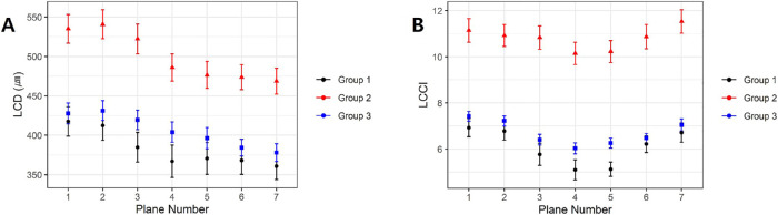
Methods: This cross-sectional study matched 24 patients diagnosed with ADOA (24 eyes) by age and retinal nerve fiber layer thickness with 48 patients diagnosed with NTG (48 eyes) by age with 48 healthy controls (48 eyes). Optic nerve heads were scanned by enhanced-depth imaging (EDI) optical coherence tomography (OCT). The LC curvature index (LCCI) and LC depth (LCD) on B-scan images obtained using EDI-OCT were measured at seven locations spaced equidistantly across the vertical optic disc diameter and compared among the NTG, ADOA, and control groups.
Results: Mean LCCI and LCD were significantly greater in NTG than in ADOA and healthy eyes (P < 0.001 each) but did not differ significantly in ADOA and healthy eyes.
Conclusions: NTG eyes have a more posteriorly curved and deeper LC than ADOA and healthy eyes. This finding provides insight into the role of LC morphology in NTG and provides a clinical clue to distinguish between NTG and ADOA.
Conflict of interest statement
Disclosure:
Figures


Similar articles
-
Differentiation of Nonarteritic Anterior Ischemic Optic Neuropathy from Normal Tension Glaucoma by Comparison of the Lamina Cribrosa.Invest Ophthalmol Vis Sci. 2020 Jul 1;61(8):21. doi: 10.1167/iovs.61.8.21. Invest Ophthalmol Vis Sci. 2020. PMID: 32668001 Free PMC article.
-
Intereye Comparison of Lamina Cribrosa Curvature in Normal Tension Glaucoma Patients With Unilateral Damage.Invest Ophthalmol Vis Sci. 2019 Jun 3;60(7):2423-2430. doi: 10.1167/iovs.19-26828. Invest Ophthalmol Vis Sci. 2019. PMID: 31158274
-
Posterior displacement of the lamina cribrosa in normal-tension and high-tension glaucoma.Acta Ophthalmol. 2016 Sep;94(6):e492-500. doi: 10.1111/aos.13012. Epub 2016 Mar 24. Acta Ophthalmol. 2016. PMID: 27009574
-
[Aiming for zero blindness].Nippon Ganka Gakkai Zasshi. 2015 Mar;119(3):168-93; discussion 194. Nippon Ganka Gakkai Zasshi. 2015. PMID: 25854109 Review. Japanese.
-
Optical Coherence Tomography Imaging of the Lamina Cribrosa: Structural Biomarkers in Nonglaucomatous Diseases.J Ophthalmol. 2021 Feb 19;2021:8844614. doi: 10.1155/2021/8844614. eCollection 2021. J Ophthalmol. 2021. PMID: 33680508 Free PMC article. Review.
Cited by
-
Morphologic Changes in the Lamina Cribrosa Upon Intraocular Pressure Lowering in Patients With Normal Tension Glaucoma.Invest Ophthalmol Vis Sci. 2022 Feb 1;63(2):23. doi: 10.1167/iovs.63.2.23. Invest Ophthalmol Vis Sci. 2022. PMID: 35147659 Free PMC article.
-
Photopic negative response recorded with RETeval system in eyes with optic nerve disorders.Sci Rep. 2022 May 31;12(1):9091. doi: 10.1038/s41598-022-12971-2. Sci Rep. 2022. PMID: 35641565 Free PMC article.
-
Visual Function and Inner Retinal Structure in Relation to Birth Factors in Autosomal Dominant Optic Atrophy.Invest Ophthalmol Vis Sci. 2023 Jul 3;64(10):32. doi: 10.1167/iovs.64.10.32. Invest Ophthalmol Vis Sci. 2023. PMID: 37498569 Free PMC article.
-
Critical Impact of Working Distance on OCT Imaging: Correction of Optical Distortion and Its Effects on Measuring Retinal Curvature.Invest Ophthalmol Vis Sci. 2024 Oct 1;65(12):10. doi: 10.1167/iovs.65.12.10. Invest Ophthalmol Vis Sci. 2024. PMID: 39374011 Free PMC article.
-
Comparing the morphology of optic nerve head and lamina cribrosa in full-term and preterm school-aged children.Eye (Lond). 2024 Jul;38(10):1964-1970. doi: 10.1038/s41433-024-03053-w. Epub 2024 Apr 17. Eye (Lond). 2024. PMID: 38632472 Free PMC article.
References
-
- O'Neill EC, Mackey DA, Connell PP, Hewitt AW, Danesh-Meyer HV, Crowston JG. The optic nerve head in hereditary optic neuropathies. Nat Rev Neurol. 2009; 5: 277–287. - PubMed
-
- Alexander C, Votruba M, Pesch UE, et al. .. OPA1, encoding a dynamin-related GTPase, is mutated in autosomal dominant optic atrophy linked to chromosome 3q28. Nat Genet. 2000; 26: 211–215. - PubMed
-
- Delettre C, Lenaers G, Griffoin JM, et al. .. Nuclear gene OPA1, encoding a mitochondrial dynamin-related protein, is mutated in dominant optic atrophy. Nat Genet. 2000; 26: 207–210. - PubMed
-
- Caldwell JB, Howard RO, Riggs LA. Dominant juvenile optic atrophy. A study in two families and review of hereditary disease in childhood. Arch Ophthalmol. 1971; 85: 133–147. - PubMed
Publication types
MeSH terms
LinkOut - more resources
Full Text Sources