. 2020 Jun 19;48(11):6108-6119.
doi: 10.1093/nar/gkaa365.

Protamine loops DNA in multiple steps

Affiliations

Protamine loops DNA in multiple steps

Obinna A Ukogu et al. Nucleic Acids Res. .

Abstract

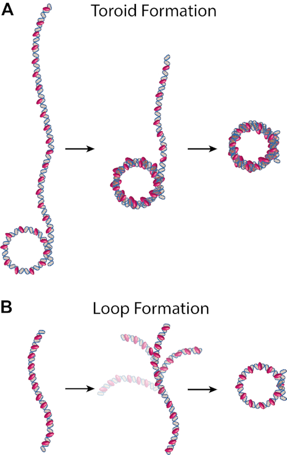
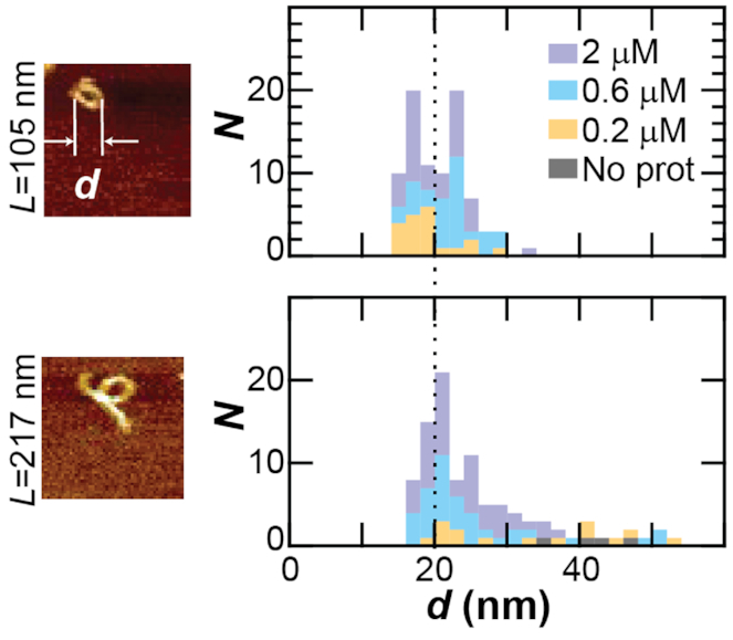
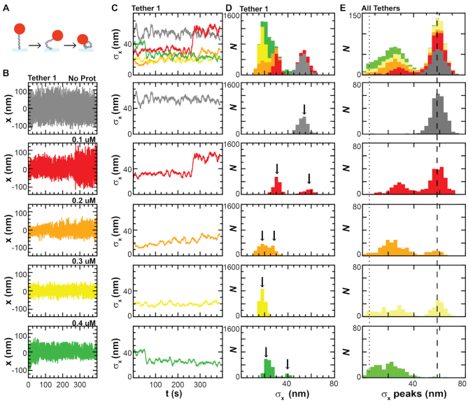
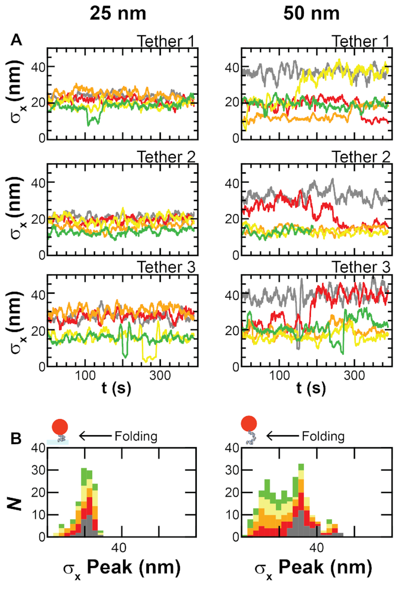
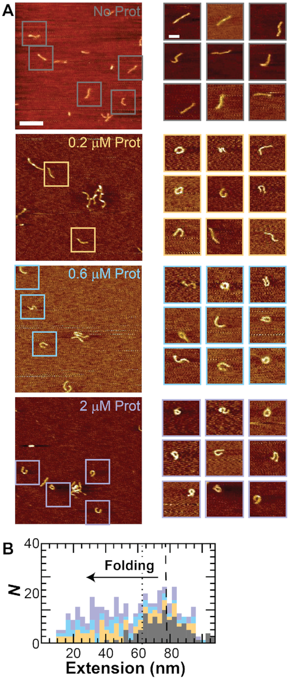
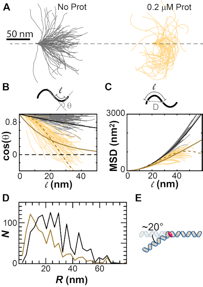
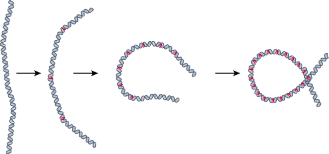
Protamine proteins dramatically condense DNA in sperm to almost crystalline packing levels. Here, we measure the first step in the in vitro pathway, the folding of DNA into a single loop. Current models for DNA loop formation are one-step, all-or-nothing models with a looped state and an unlooped state. However, when we use a Tethered Particle Motion (TPM) assay to measure the dynamic, real-time looping of DNA by protamine, we observe the presence of multiple folded states that are long-lived (∼100 s) and reversible. In addition, we measure folding on DNA molecules that are too short to form loops. This suggests that protamine is using a multi-step process to loop the DNA rather than a one-step process. To visualize the DNA structures, we used an Atomic Force Microscopy (AFM) assay. We see that some folded DNA molecules are loops with a ∼10-nm radius and some of the folded molecules are partial loops-c-shapes or s-shapes-that have a radius of curvature of ∼10 nm. Further analysis of these structures suggest that protamine is bending the DNA to achieve this curvature rather than increasing the flexibility of the DNA. We therefore conclude that protamine loops DNA in multiple steps, bending it into a loop.


Figures

Figure 1.
Figure 1.
Models for toroid and loop formation. (A) A toroid formation model. Multiple protamine molecules (pink) bind the DNA and form a single loop. Subsequent loops stack one by one on top of the first loop, forming the toroid. (B) One-step loop formation model. Multiple protamine molecules bind the DNA. Random fluctuations in the conformation of the DNA then create opportunities for one or multiple protamine molecules to stabilize the location where the DNA strands cross, forming a loop in one step. Not to scale. Numbers of protamine molecules and binding locations could vary from what is shown.
Figure 2.
Figure 2.
DNA loops folded by protamine have ∼20 nm diameters. (Left) Images of DNA loops folded by protamine. DNA length, L, is either 105 nm or 217 nm. Computer algorithm calculates the loop diameter d (see Materials and Methods). Width of images is 200 nm. (Right) Histograms of the loop diameter at different protamine concentrations (gray = no protamine, yellow = 0.2 μM, blue = 0.6 μM, and purple = 2 μM protamine). Histograms are stacked. Mean loop diameter for 105-nm-length DNA is 20 nm (dotted line).
Figure 3.
Figure 3.
Loop formation is multi-step with long-lived, reversible states. (A) Assay consists of particle tethered to the surface via a DNA molecule. As protamine loops the DNA, the tether shortens, decreasing particle motion. (B) Particle position, x, at 5 Hz for a single tether without protamine (gray) and with protamine titrations of 0.1 μM (red), 0.2 μM (orange), 0.3 μM (yellow), and 0.4 μM (green). (C) Rolling standard deviation, σx, at 0.1 Hz of the x traces at each concentration plotted together (top) and individually (bottom). Traces of σx decrease as the protamine concentration increases and the DNA folds. (D) Histograms of the σx traces at each concentration stacked together (top) and plotted individually (bottom) show the presence of multiple peaks (black arrows), indicating multiple folded states. The height of the peak is equal to the number of frames in the state, with many states lasting longer than 100 s. (E) We repeat the measurement made in B-D on all the tethers (N = 103) and determine the peaks in the σx histogram (σx peaks). We then histogram the σx peaks for all tethers at each concentration, stacking them together (top) and plotting individually (bottom). The unfolded state has a mean σx of 59 ± 5 nm (dashed line). The detection limit (dotted line) of the instrument is 6 nm.
Figure 4.
Figure 4.
DNA folding occurs even at short DNA lengths. (A) Plots of the rolling standard deviation, σx, at each protamine concentration over time for three individual 25-nm-length DNA tethers and three individual 50-nm-length DNA tethers. A decrease in σx indicates DNA folding. Data is at 0.1 Hz. (B) Histograms of σx peaks for all of the 25-nm-length and 50-nm-length DNA tethers. Histograms at each protamine concentration are stacked one on top of the other. Color scheme is the same as Figure 2.
Figure 5.
Figure 5.
Intermediate structures are partial loops. (A) (Left) AFM images of DNA (L = 105 nm) without protamine (gray) and with 0.2 μM (orange), 0.6 μM (blue), or 2 μM (purple) protamine show individual molecules (box). Scale bar is 200 nm. (Right) Representative individual molecules show unfolded and folded structures. The folded structures are loops and partial loops—c-shapes and s-shapes. Scale bar is 50 nm. (B) Computer algorithm calculates the end-to-end extension (inset) for all molecules that are not loops. Plot shows stacked histograms of the extension. Color scheme same as in A. Unfolded molecules have a mean extension of 77 nm (dashed line). Molecules that are in an intermediate folding state are defined as having a fractional extension of <0.6 (dotted line).
Figure 6.
Figure 6.
Intermediate folding states have a radius of curvature of ∼10 nm. We compare DNA molecules without protamine (gray) to molecules with 0.2 μM protamine that are folded but not looped (yellow). (A) Contours for the molecules are aligned so that they start at the origin and initially have tangent vectors that point along the x-axis. (B) The cosine of the angle, θ, between segments along the contour decreases with contour length, . Fits using the model of the flexible polymer (bold, solid lines) approach zero. The fit using the model of the contour bending along a circle (dashed line) crosses over zero and becomes negative. (C) The mean-squared displacement (MSD) calculated from the displacement D for points along the DNA contour as a function of contour length. Fits using the model of the flexible polymer (bold, solid lines) increase with contour length. The fit using the model of the contour bending along a circle (dashed line) decreases at high (>40 nm) contour lengths. (D) Histograms of the radius of curvature for molecules in 0.2 μM protamine that are folded but not looped (dark yellow) and molecules without protamine (black). (E) Cartoon of single protamine creating a 20° bend in the DNA.
Figure 7.
Figure 7.
Multi-step looping model. Protamine (pink) loops the DNA in multiple steps. Each step is an enthalpic bending of the DNA along a circular path. Multiple steps produce a loop. Bending could be carried out by one or more protamine molecules. Not to scale.

References

    1. Pogany G.C., Corzett M., Weston S., Balhorn R.. DNA and protein content of mouse sperm. implications regarding sperm chromatin structure. Exp. Cell Res. 1981; 136:127–136. - PubMed
    1. Hud N.V., Milanovich F.P., Balhorn R.. Evidence of novel secondary structure in DNA-bound protamine is revealed by Raman spectroscopy. Biochemistry. 1994; 33:7528–7535. - PubMed
    1. Teif V.B., Bohinc K.. Condensed DNA: condensing the concepts. Prog. Biophys. Mol. Biol. 2011; 105:208–222. - PubMed
    1. Sotolongo B., Lino E., Ward W.S.. Ability of hamster spermatozoa to digest their own DNA. Biol. Reprod. 2003; 69:2029–2035. - PubMed
    1. Balhorn R. The protamine family of sperm nuclear proteins. Genome Biol. 2007; 8:227. - PMC - PubMed

Publication types