Review
. 2020 Sep 29;7(1):121-141.
doi: 10.1146/annurev-virology-010320-052547. Epub 2020 May 11.

Ecology, Structure, and Evolution of Shigella Phages

Affiliations
Review

Ecology, Structure, and Evolution of Shigella Phages

Sundharraman Subramanian et al. Annu Rev Virol. .

Abstract

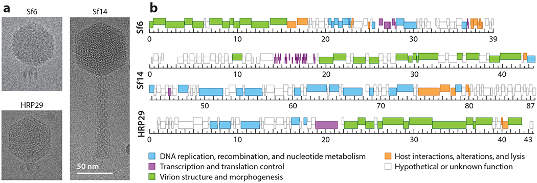
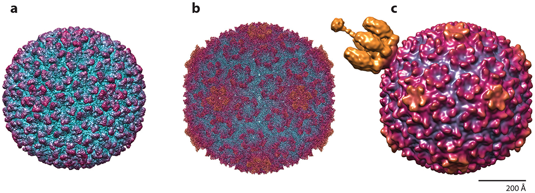
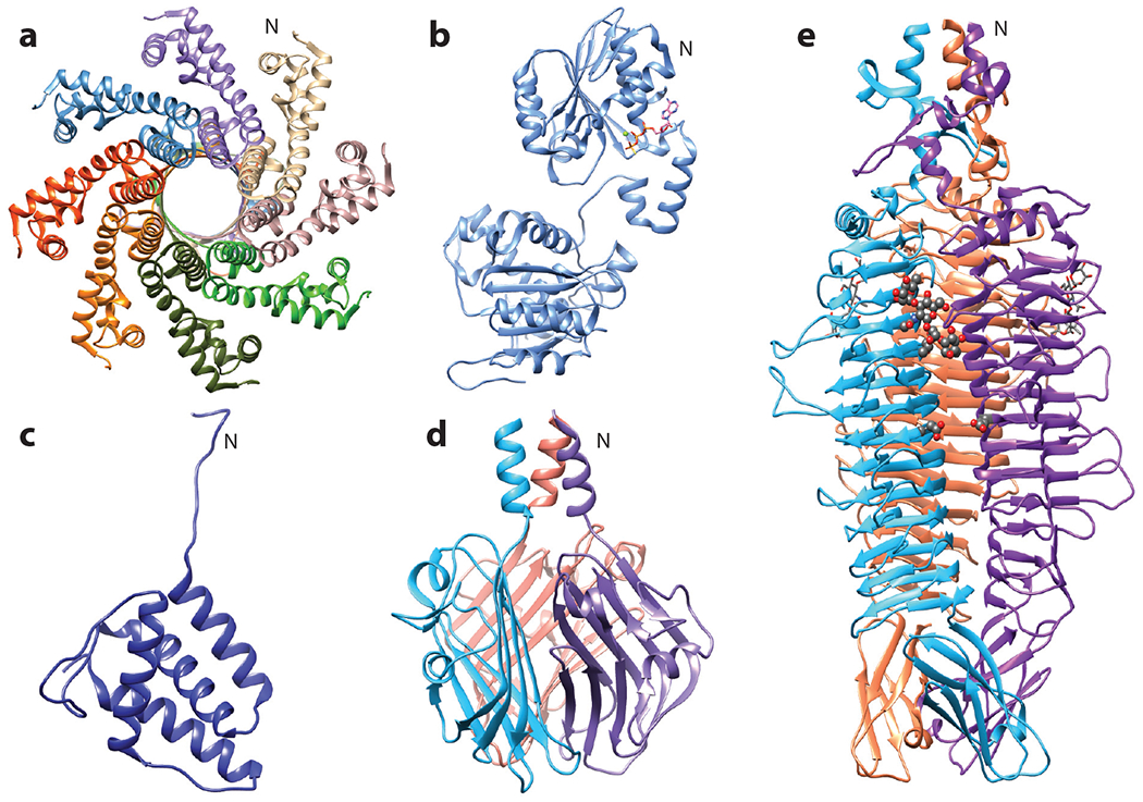
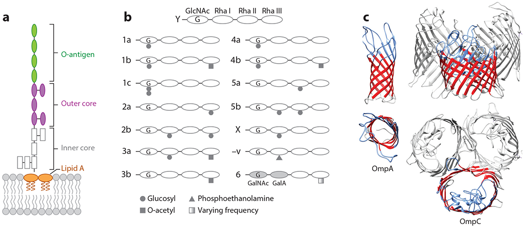
Numerous bacteriophages-viruses of bacteria, also known as phages-have been described for hundreds of bacterial species. The Gram-negative Shigella species are close relatives of Escherichia coli, yet relatively few previously described phages appear to exclusively infect this genus. Recent efforts to isolate Shigella phages have indicated these viruses are surprisingly abundant in the environment and have distinct genomic and structural properties. In addition, at least one model system used for experimental evolution studies has revealed a unique mechanism for developing faster infection cycles. Differences between these bacteriophages and other well-described model systems may mirror differences between their hosts' ecology and defense mechanisms. In this review, we discuss the history of Shigella phages and recent developments in their isolation and characterization and the structural information available for three model systems, Sf6, Sf14, and HRP29; we also provide an overview of potential selective pressures guiding both Shigella phage and host evolution.

Keywords: Omps; Shigella; lipopolysaccharide; myoviruses; phage biology; podoviruses.


Figures

Figure 1
Figure 1
Characteristics of the developing Shigella phage model systems Sf14 and HRP29 compared with the established model system Sf6. (a) Representative images from cryo-electron micrographs. (b) Genome maps, with genes colored according to function, as indicated. The ruler is in kilo-base pairs. The cluster of short, dark-colored bars at 14–18 kbp in the Sf14 genome represents transfer RNA genes.
Figure 2
Figure 2
Structures of Sf6 macromolecular complexes, colored according to radial distance. (a) Procapsid at 7.8 Å, EMDB 5724. (b) Capsid at 2.9 Å, EMDB 8314. (c) Virion at 16.0 Å, EMDB 5730. Like other precursor procapsids, it has the smallest diameter and the hexons are skewed with twofold symmetry. Upon expansion, the capsid walls become thinner, the hexons isomerize, and the internal volume increases by ~10%. The mature virion includes the tail apparatus, shown in gold, attached to the portal vertex.
Figure 3
Figure 3
Structures of Sf6 proteins. (a) Small terminase octamer at 1.65 Å, with the N terminus forming the body facing the viewer and the neck facing away, PDB 3HEF. (b) Large terminase monomer bound to ATPγS at 1.89 Å, PDB 4IEE. (c) Tail adaptor monomer at 1.77 Å, PDB 5VGT. (d) Tail needle knob trimer showing the jellyroll fold at 1 Å, PDB 3RWN. (e) Tailspike trimer with one tetrasaccharide molecule and the catalytic active site residues Asp 399 and Glu 366 shown in gray ball-stick model, resolved to 2 Å, PDB 2VBM. For multimeric proteins, individual subunits are shown in different colors. The N indicates the N terminus for one monomer.
Figure 4
Figure 4
Primary and secondary phage receptors in Shigella flexneri. Primary receptors: (a) simplified diagram of smooth lipopolysaccharide, with (b) illustrating modifications to the O-antigen, producing serotypes indicated to the left. Secondary receptors: (c) outer membrane proteins (Omps) A and C, PDB 3NB3 and 2J1N, respectively.

Similar articles

Cited by

References

    1. Pupo GM, Lan R, Reeves PR. 2000. Multiple independent origins of Shigella clones of Escherichia coli and convergent evolution of many of their characteristics. PNAS 97:10567–72 - PMC - PubMed
    1. Zhang Y, Lin K.2012. A phylogenomic analysis of Escherichia coli/Shigella group: implications of genomic features associated with pathogenicity and ecological adaptation. BMC Evol. Biol. 12:174. - PMC - PubMed
    1. Freed NE, Bumann D, Silander OK. 2016. Combining Shigella Tn-seq data with gold-standard E. coli gene deletion data suggests rare transitions between essential and non-essential gene functionality. BMC Microbiol. 16:203. - PMC - PubMed
    1. Rautureau GJP, Palama TL, Canard I, Mirande C, Chatellier S,et al. 2019. Discrimination of Escherichia coli and Shigella spp. by nuclear magnetic resonance based metabolomic characterization of culture media. ACS Infect. Dis. 5:1879–86 - PubMed
    1. Goodridge LD. 2013. Bacteriophages for managing Shigella in various clinical and non-clinical settings. Bacteriophage 3:e25098. - PMC - PubMed

LinkOut - more resources