Review
. 2020 May 7;8(2):43.
doi: 10.3390/dj8020043.

Utility of Photodynamic Therapy in Dentistry: Current Concepts

Affiliations
Review

Utility of Photodynamic Therapy in Dentistry: Current Concepts

Anette Stájer et al. Dent J (Basel). .

Abstract

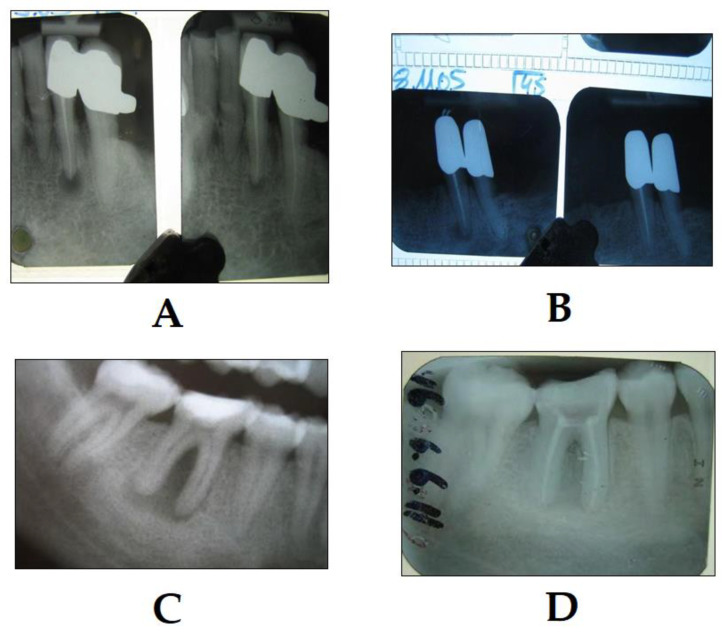
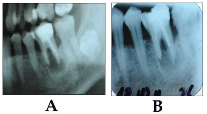
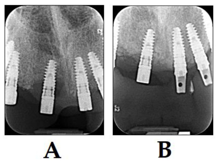
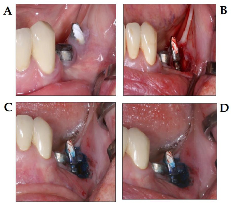
The significant growth in scientific and technological advancements within the field of dentistry has resulted in a wide range of novel treatment modalities for dentists to use. Photodynamic therapy (PDT) is an emerging, non-invasive treatment method, involving photosensitizers, light of a specific wavelength and the generation of singlet oxygen and reactive oxygen species (ROS) to eliminate unwanted eukaryotic cells (e.g., malignancies in the oral cavity) or pathogenic microorganisms. The aim of this review article is to summarize the history, general concepts, advantages and disadvantages of PDT and to provide examples for current indications of PDT in various subspecialties of dentistry (oral and maxillofacial surgery, oral medicine, endodontics, preventive dentistry, periodontology and implantology), in addition to presenting some images from our own experiences about the clinical success with PDT.

Keywords: aminolevulinic acid; caries; endodontics; implantology; oral medicine; periodontology; photodynamic therapy; photosensitizer; reactive oxygen species.


Conflict of interest statement

The authors declare no conflict of interest, monetary or otherwise.

Figures

Figure 1
Figure 1
Examples of clinically relevant photosensitizer (PS) compounds. (A) Photofrin® (hematoporphyrin or dihematoporphyrin ether); (B) Visudyne® (verteporfin); (C) 5-aminolevulinic acid (ALA); (D) talaporfin sodium (LS11); and (E) Foscan® (temoporfin).
Figure 2
Figure 2
A diagram, illustrating the various changes in energy levels, which provide the physical and molecular basis for photodynamic therapy (PDT) (adapted from [25]). 1O2: singlet oxygen, 3O2: triplet oxygen, A = absorption of photons, F = fluorescence (emission), P = phosphorescence S = singlet state, T = triplet state, IC = internal conversion, ISC = intersystem crossing, EET = energy transfer through excitation, and ET = transfer of electrons, R = substrate.
Figure 3
Figure 3
PDT of lingual candidiasis (photo courtesy of R. Szabó DMD, J. Arentz DMD and T. Nave DMD). (A) C. albicans infection of the tongue before PDT treatment; (B) application of PS compound; (C) irradiation; and (D) C. albicans infection of the tongue 36 h after PDT treatment, showing remission.
Figure 4
Figure 4
Example of PDT in endodontic treatment (photo courtesy of R. Szabó DMD, J. Arentz DMD and T. Nave DMD). (A) Extracted lower incisor after chemo-mechanical preparation and application of the PS into the canal; (B) penetration of the PS into the canal, 7 h after application, the PS already shows penetration into the apical region and lateral canals; and (C) treatment with laser light; the PDT light system is able to align the laser light in a way to allow for the activation of as many as possible PSs inside the canal to achieve the optimum effect. Following this step, root canals may be obturated.
Figure 5
Figure 5
Success of PDT in the treatment of chronic periapical periodontitis (photo courtesy of R. Szabó DMD, J. Arentz DMD and T. Nave DMD). (A) The X-ray on the left shows the condition immediately after PDT, and the X-ray on the right shows the condition 4 months later; note the significant decrease in the size of the periapical lesion; (B) the X-ray on the left shows the condition immediately after PDT, and the X-ray on the right shows the condition 12 months later; note the significant decrease in the size of the periapical lesion. The same case is depicted in a lower molar tooth with chronic periodontitis, where (C) shows the condition immediately after the endodontic treatment and PDT, and (D) shows the condition after 3 months after the endodontic treatment, PDT therapy and root canal obturation; note the significant decrease in the size of the periapical lesion.
Figure 6
Figure 6
Example of PDT in periodontal therapy (photo courtesy of R. Szabó DMD, J. Arentz DMD and T. Nave DMD). (A) X-ray shows a perio-endo lesion of a tooth before PDT; and (B) X-ray shows the condition three months after PDT. Bone regeneration is visible showing clear signs of clinical improvement.
Figure 7
Figure 7
Treatment of patients with peri-implantitis with PDT and BIOOSS bone graft material (photo courtesy of R. Szabó DMD, J. Arentz DMD and T. Nave DMD). (A) X-ray of diagnosis of peri-implantitis before treatment; and (B) X-ray 3 months after treatment with PDT and BIOOSS bone graft material. Bone regeneration is visible showing a clear sign of healing.
Figure 8
Figure 8
PDT in the treatment of periimplantitis, using the Photolase® PDT system (photo courtesy of Z. Baráth DMD PhD). (A) Initial stage peri-implantitis; (B) surgical flap elevation; (C) application of photosensitizer after surgical debridement; and (D) conditions directly after the diode laser therapy.

References

    1. Slavkin H.C. Evolution of the scientific basis for dentistry and its impact on dental education: Past, present, and future. J. Dent. Educ. 2012;76:28–35. - PubMed
    1. Polverini P.J., Krebsbach P.H. Research and Discovery Science and the Future of Dental Education and Practice. J. Dent. Educ. 2017;81:eS97–eS107. doi: 10.21815/JDE.017.040. - DOI - PubMed
    1. Gutmann J.L. The maturation of science within dentistry: The impact of critical milestones and visionary leaders on contemporary achievements. J. Hist. Dent. 2009;57:109–122. - PubMed
    1. Issa M.C.A., Manela-Azulay M. Photodynamic therapy: A review of the literature and image documentation. An. Bras. Dermatol. 2010;85:501–511. doi: 10.1590/S0365-05962010000400011. - DOI - PubMed
    1. Wan M.T., Lin J.Y. Current evidence and applications of photodynamic therapy in dermatology. Clin. Cosmet. Investig. Dermatol. 2014;7:145–163. - PMC - PubMed

LinkOut - more resources