

Walnut (Juglans regia L.) Septum: Assessment of Bioactive Molecules and In Vitro Biological Effects
- PMID: 32392837
- PMCID: PMC7248768
- DOI: 10.3390/molecules25092187
Walnut (Juglans regia L.) Septum: Assessment of Bioactive Molecules and In Vitro Biological Effects
Abstract
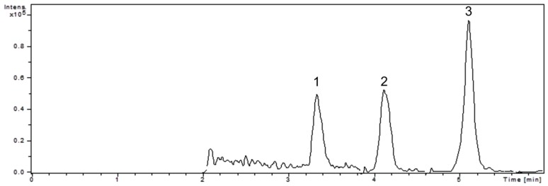
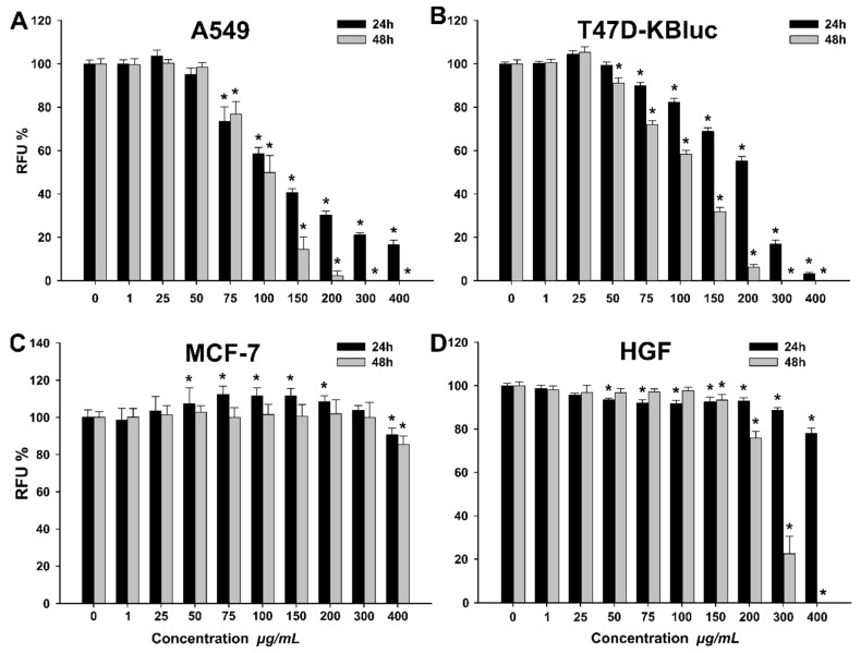
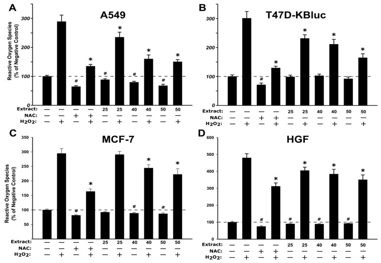
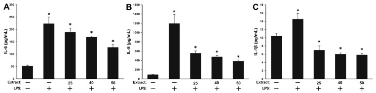
Walnut (Juglans regia L.) septum represents an interesting bioactive compound source by-product. In our study, a rich phenolic walnut septum extract, previously selected, was further examined. The tocopherol content determined by liquid chromatography-tandem mass spectrometry (LC-MS/MS) revealed higher amounts of α-tocopherol compared to γ- and δ-tocopherols. Moreover, several biological activities were investigated. The in vitro inhibiting assessment against acetylcholinesterase, α-glucosidase, or lipase attested a real management potential in diabetes or obesity. The extract demonstrated very strong antimicrobial potential against Staphylococcus aureus, Pseudomonas aeruginosa and Salmonella enteritidis. It also revealed moderate (36.08%) and strong (43.27%) antimutagenic inhibitory effects against TA 98 and TA 100 strains. The cytotoxicity of the extract was assessed on cancerous (A549, T47D-KBluc, MCF-7) and normal (human gingival fibroblasts (HGF)) cell lines. Flow cytometry measurements confirmed the cytotoxicity of the extract in the cancerous cell lines. Additionally, the extract demonstrated antioxidant activity on all four cell types, as well as anti-inflammatory activity by lowering the inflammatory cytokines (interleukin-6 (IL-6), interleukin-8 (IL-8), interleukin-1 β (IL-1β)) evaluated in HGF cells. To the best of our knowledge, most of the cellular model analyses were performed for the first time in this matrix. The results prove that walnut septum may be a potential phytochemical source for pharmaceutical and food industry.
Keywords: LC-MS/MS; anti-inflammatory activity; antimicrobial; antimutagenic; antioxidant; by-product; enzyme inhibitor; tocopherols; walnut septum.
Conflict of interest statement
The authors declare that they have no known competing financial interests or personal relationships that could have appeared to influence the work reported in this paper.
Figures
References
-
- Vodnar D.C., Călinoiu L.F., Dulf F.V., Ştefănescu E., Crişan G., Socaciu C. Identification of the bioactive compounds and antioxidant, antimutagenic and antimicrobial activities of thermally processed agro-industrial waste. Food Chem. 2017;231:131–140. doi: 10.1016/j.foodchem.2017.03.131. - DOI - PubMed
-
- Santos A., Barros L., Calhelha R.C., Dueñas M., Carvalho A.M., Santos-Buelga C., Ferreira I.C.F.R. Leaves and decoction of Juglans regia L.: Different performances regarding bioactive compounds and in vitro antioxidant and antitumor effects. Ind Crop. Prod. 2013;51:430–436. doi: 10.1016/j.indcrop.2013.10.003. - DOI
-
- Rusu M.E., Gheldiu A.-M., Mocan A., Moldovan C., Popa D.-S., Tomuta I., Vlase L. Process Optimization for Improved Phenolic Compounds Recovery from Walnut (Juglans regia L.) Septum: Phytochemical Profile and Biological Activities. Molecules. 2018;23:2814. doi: 10.3390/molecules23112814. - DOI - PMC - PubMed
MeSH terms
Substances
LinkOut - more resources
Full Text Sources
Medical
Molecular Biology Databases
