

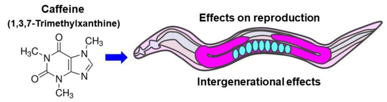
Maternal Caffeine Intake Disrupts Eggshell Integrity and Retards Larval Development by Reducing Yolk Production in a Caenorhabditis elegans Model
- PMID: 32392893
- PMCID: PMC7284833
- DOI: 10.3390/nu12051334
Maternal Caffeine Intake Disrupts Eggshell Integrity and Retards Larval Development by Reducing Yolk Production in a Caenorhabditis elegans Model
Abstract
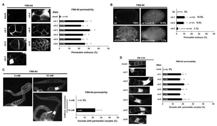
During pregnancy, most women are exposed to caffeine, which is a widely consumed psychoactive substance. However, the consequences of maternal caffeine intake on the child remain largely unknown. Here, we investigated the intergenerational effects of maternal caffeine intake on offspring in a Caenorhabditis elegans model. We treated a young mother (P0) with 10 mM of caffeine equivalent to 2-5 cans of commercial energy drinks and examined its reproduction and growth rate from P0 to F2 generation. The fertility decreased and embryonic lethality increased by defective oocytes and eggshell integrity in caffeine-ingested mothers, and F1 larval development severely retarded. These results were due to decreased production of vitellogenin protein (yolk) in caffeine-ingested mothers. Furthermore, effects of RNA interference of vitellogenin (vit) genes, vit-1 to vit-6, in P0 mothers can mimic those by caffeine-ingested mothers. In addition, RNA interference (RNAi) depletion of unc-62 (human Meis homeobox), a transcriptional activator for vit genes, also showed similar effects induced by caffeine intake. Taken together, maternal caffeine intake reduced yolk production mediated by the UNC-62 transcription factor, thereby disrupting oocyte and eggshell integrity and retarding larval development. Our study suggests the clinical significance of caffeine intake for prospective mothers.
Keywords: 1,3,7-trimethylxanthine; Caenorhabditis elegans; UNC-62; caffeine; eggshell integrity; intergenerational effect; maternal effect; reproduction; vitellogenin; yolk protein.
Conflict of interest statement
The authors declare no conflicts of interest.
Figures


Similar articles
-
Caffeine-Induced Upregulation of pas-1 and pas-3 Enhances Intestinal Integrity by Reducing Vitellogenin in Aged Caenorhabditis elegans Model.Nutrients. 2024 Dec 12;16(24):4298. doi: 10.3390/nu16244298. Nutrients. 2024. PMID: 39770921 Free PMC article.
-
Caffeine induces high expression of cyp-35A family genes and inhibits the early larval development in Caenorhabditis elegans.Mol Cells. 2015 Mar;38(3):236-42. doi: 10.14348/molcells.2015.2282. Epub 2015 Jan 16. Mol Cells. 2015. PMID: 25591395 Free PMC article.
-
Reversible reprotoxic effects of manganese through DAF-16 transcription factor activation and vitellogenin downregulation in Caenorhabditis elegans.Life Sci. 2016 Apr 15;151:218-223. doi: 10.1016/j.lfs.2016.03.016. Epub 2016 Mar 10. Life Sci. 2016. PMID: 26972607
-
[Effects of coffee and caffeine on fertility, reproduction, lactation, and development. Review of human and animal data].J Gynecol Obstet Biol Reprod (Paris). 1994;23(3):241-56. J Gynecol Obstet Biol Reprod (Paris). 1994. PMID: 8051344 Review. French.
-
Vitellogenins - Yolk Gene Function and Regulation in Caenorhabditis elegans.Front Physiol. 2019 Aug 21;10:1067. doi: 10.3389/fphys.2019.01067. eCollection 2019. Front Physiol. 2019. PMID: 31551797 Free PMC article. Review.
Cited by
-
Vitamin B12 Supplementation Improves Oocyte Development by Modulating Mitochondria and Yolk Protein in a Caffeine-Ingested Caenorhabditis elegans Model.Antioxidants (Basel). 2023 Dec 28;13(1):53. doi: 10.3390/antiox13010053. Antioxidants (Basel). 2023. PMID: 38247478 Free PMC article.
-
A role for worm cutl-24 in background- and parent-of-origin-dependent ER stress resistance.BMC Genomics. 2022 Dec 20;23(1):842. doi: 10.1186/s12864-022-09063-w. BMC Genomics. 2022. PMID: 36539699 Free PMC article.
-
Maternal Gliadin Intake Reduces Oocyte Quality with Chromosomal Aberrations and Increases Embryonic Lethality through Oxidative Stress in a Caenorhabditis elegans Model.Nutrients. 2022 Dec 19;14(24):5403. doi: 10.3390/nu14245403. Nutrients. 2022. PMID: 36558561 Free PMC article.
-
Molecular Characterization of the Von Willebrand Factor Type D Domain of Vitellogenin from Takifugu flavidus.Mar Drugs. 2021 Mar 25;19(4):181. doi: 10.3390/md19040181. Mar Drugs. 2021. PMID: 33806251 Free PMC article.
-
Effects of Phosphoethanolamine Supplementation on Mitochondrial Activity and Lipogenesis in a Caffeine Ingestion Caenorhabditis elegans Model.Nutrients. 2020 Oct 30;12(11):3348. doi: 10.3390/nu12113348. Nutrients. 2020. PMID: 33143181 Free PMC article.
References
-
- Papadopoulou E., Botton J., Brantsæter A.L., Haugen M., Alexander J., Meltzer H.M., Bacelis J., Elfvin A., Jacobsson B., Sengpiel V. Maternal caffeine intake during pregnancy and childhood growth and overweight: Results from a large Norwegian prospective observational cohort study. BMJ Open. 2018;8:e018895. doi: 10.1136/bmjopen-2017-018895. - DOI - PMC - PubMed
-
- Calvo D.R., Martorell P., Genovés S., Gosálbez L. Development of novel functional ingredients: Need for testing systems and solutions with Caenorhabditis elegans. Trends Food Sci. Technol. 2016;54:197–203. doi: 10.1016/j.tifs.2016.05.006. - DOI
MeSH terms
Substances
Grants and funding
LinkOut - more resources
Full Text Sources
Medical
Research Materials
Miscellaneous
