

Intravitreal safety profiles of sol-gel mesoporous silica microparticles and the degradation product (Si(OH)4)
- PMID: 32393079
- PMCID: PMC7269085
- DOI: 10.1080/10717544.2020.1760401
Intravitreal safety profiles of sol-gel mesoporous silica microparticles and the degradation product (Si(OH)4)
Abstract
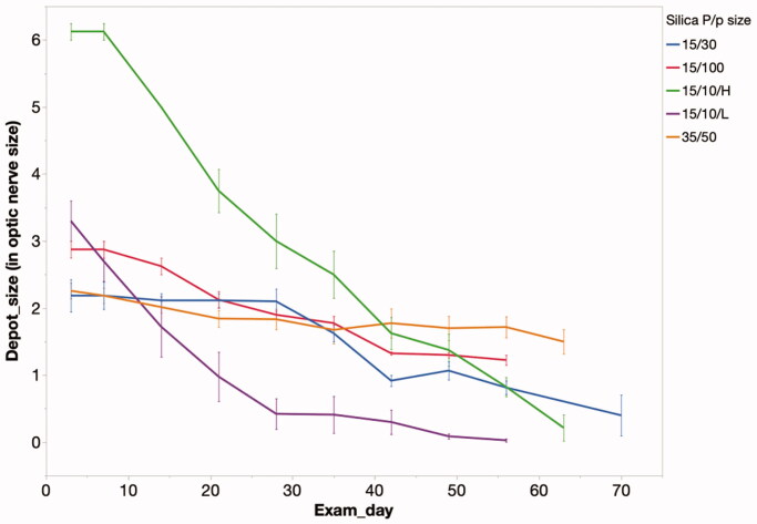
Mesoporous silica has attracted significant attention in the drug delivery area; however, impurities can be a source of toxicity. The current study used commercial microparticles produced at large scale in a well-controlled environment. Micrometer sized mesoporous silica particles were acquired through a commercial vendor and pore structures were characterized by SEM. The three silica particle formulations had a diameter of 15 micrometers and three different pore sizes of 10 nm, 30 nm, and 100 nm. The fourth formulation had particle size of 20-40 micrometers with 50 nm pores. Before in vivo tests, an in vitro cytotoxicity test was conducted with silicic acid, derived from the sol-gel particles, on EA.hy926 cells. Low concentration (2.5 µg/mL) of silicic acid showed no cytotoxicity; however, high concentration (25 µg/mL) was cytotoxic. In vivo intravitreal injection demonstrated that 15 um silica particles with 10 nm pore were safe in both rabbit and guinea pig eyes and the particles lasted in the vitreous for longer than two months. Formulations of with larger pores demonstrated variable localized vitreous cloudiness around the sol-gel particle depot and mild inflammatory cells in the aqueous humor. The incidence of reaction trended higher with larger pores (10 nm: 0%, 30 nm: 29%, 50 nm: 71%, 100 nm: 100%, p < .0001, Cochran Armitage Trend Test). Sol-gel mesoporous silica particles have uniform particle sizes and well-defined pores, which is an advantage for implantation via a fine needle. Selected formulations may be used as an intraocular drug delivery system with proper loading and encapsulation.
Keywords: Intravitreal drug delivery; rabbit and guinea pig eyes; silica pore size and ocular toxicity; silicic acid cytotoxicity; sol-gel mesoporous silica.
Figures


Similar articles
-
Evaluation of the Ocular Safety of Hollow Mesoporous Organosilica Nanoparticles with Different Tetrasulfur Bond Content.Int J Nanomedicine. 2024 Jul 16;19:7123-7136. doi: 10.2147/IJN.S464524. eCollection 2024. Int J Nanomedicine. 2024. PMID: 39055375 Free PMC article.
-
A Biodegradation Study of SBA-15 Microparticles in Simulated Body Fluid and in Vivo.Langmuir. 2015 Jun 16;31(23):6457-62. doi: 10.1021/acs.langmuir.5b01316. Epub 2015 Jun 4. Langmuir. 2015. PMID: 26013363
-
Synthesis of mesoporous silica-gel core-shell structural microparticles and their multiple drug delivery.Drug Deliv. 2015 Jan;22(1):69-78. doi: 10.3109/10717544.2013.859768. Epub 2013 Nov 25. Drug Deliv. 2015. PMID: 24266606
-
Engineering silica particles as oral drug delivery vehicles.Curr Pharm Des. 2008;14(18):1821-31. doi: 10.2174/138161208784746671. Curr Pharm Des. 2008. PMID: 18673185 Review.
-
Mesoporous silica formulation strategies for drug dissolution enhancement: a review.Expert Opin Drug Deliv. 2016;13(1):93-108. doi: 10.1517/17425247.2016.1100165. Epub 2015 Nov 7. Expert Opin Drug Deliv. 2016. PMID: 26549623 Review.
Cited by
-
PLGA-modified Syloid®-based microparticles for the ocular delivery of terconazole: in-vitro and in-vivo investigations.Drug Deliv. 2022 Dec;29(1):2117-2129. doi: 10.1080/10717544.2022.2092239. Drug Deliv. 2022. PMID: 35838555 Free PMC article.
-
Single subconjunctival injection formulation using sol-gel mesoporous silica as a controlled release system for drop-free post-cataract surgery care.J Cataract Refract Surg. 2020 Nov;46(11):1548-1553. doi: 10.1097/j.jcrs.0000000000000366. J Cataract Refract Surg. 2020. PMID: 32818352 Free PMC article.
-
Cyclodextrin Stabilized Freeze-Dried Silica/Chitosan Nanoparticles for Improved Terconazole Ocular Bioavailability.Pharmaceutics. 2022 Feb 22;14(3):470. doi: 10.3390/pharmaceutics14030470. Pharmaceutics. 2022. PMID: 35335847 Free PMC article.
-
Drug Delivery Challenges and Current Progress in Nanocarrier-Based Ocular Therapeutic System.Gels. 2022 Jan 28;8(2):82. doi: 10.3390/gels8020082. Gels. 2022. PMID: 35200463 Free PMC article. Review.
References
-
- Bae Y, Lee S, Kim SH. (2011). Chrysin suppresses mast cell-mediated allergic inflammation: involvement of calcium, caspase-1 and nuclear factor-kappaB. Toxicol Appl Pharmacol 254:56–64. - PubMed
-
- Bellocq NC, Pun SH, Jensen GS, Davis ME. (2003). Transferrin-containing, cyclodextrin polymer-based particles for tumor-targeted gene delivery. Bioconjugate Chem 14:1122–32. - PubMed
-
- Bimbo LM, Sarparanta M, Santos HA, et al. (2010). Biocompatibility of thermally hydrocarbonized porous silicon nanoparticles and their biodistribution in rats. Acs Nano 4:3023–32. - PubMed