. 2020 Nov 11;20(2):89-103.
doi: 10.3727/105221620X15889714507961. Epub 2020 May 11.

Downregulation of p16 Decreases Biliary Damage and Liver Fibrosis in the Mdr2/ Mouse Model of Primary Sclerosing Cholangitis

Affiliations

Downregulation of p16 Decreases Biliary Damage and Liver Fibrosis in the Mdr2/ Mouse Model of Primary Sclerosing Cholangitis

Konstantina Kyritsi et al. Gene Expr. .

Abstract

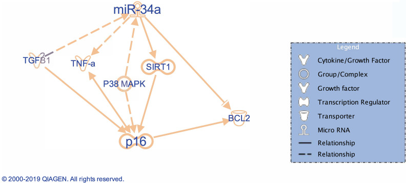
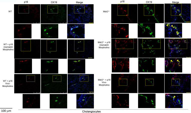
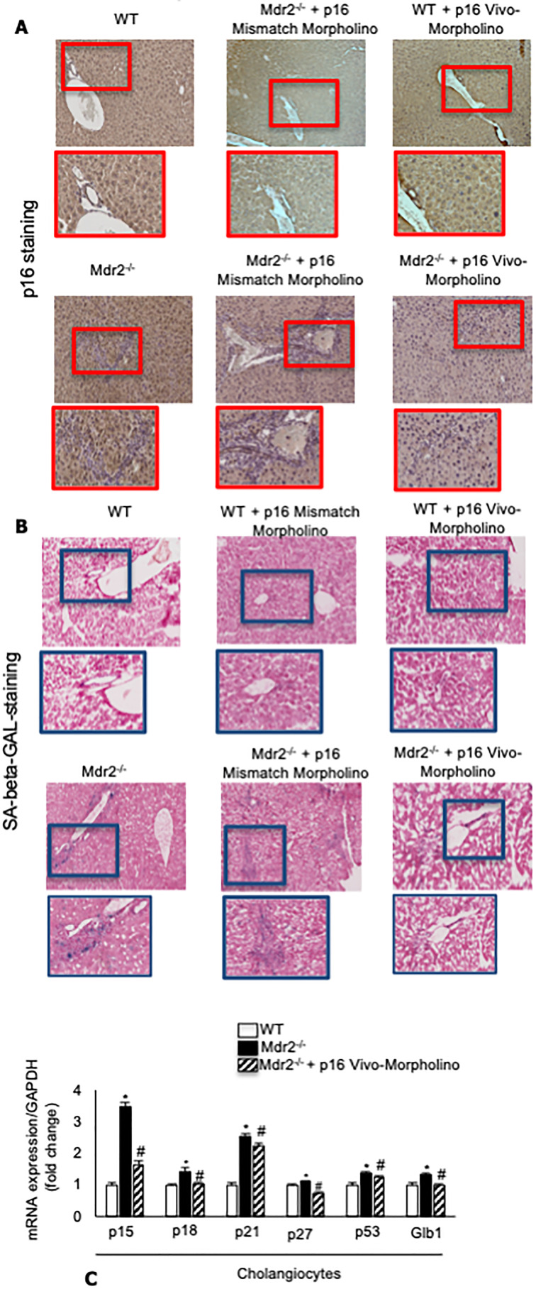
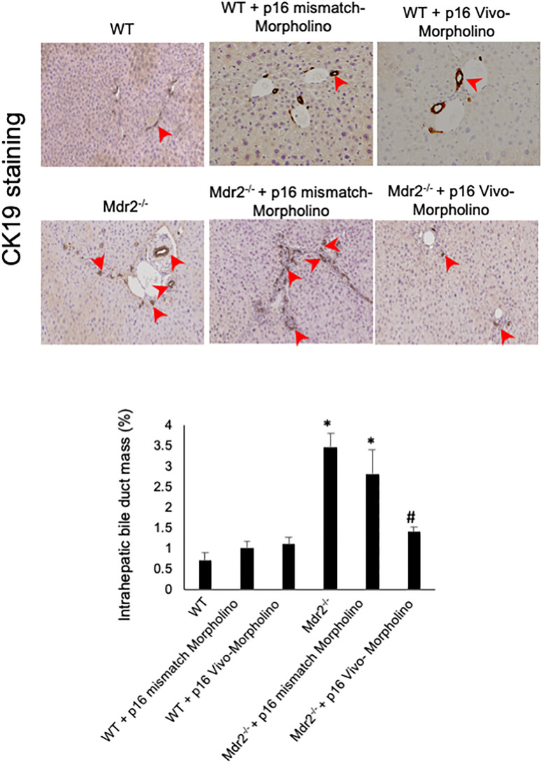
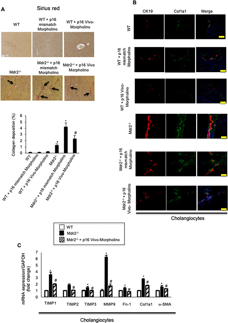
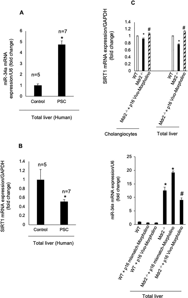
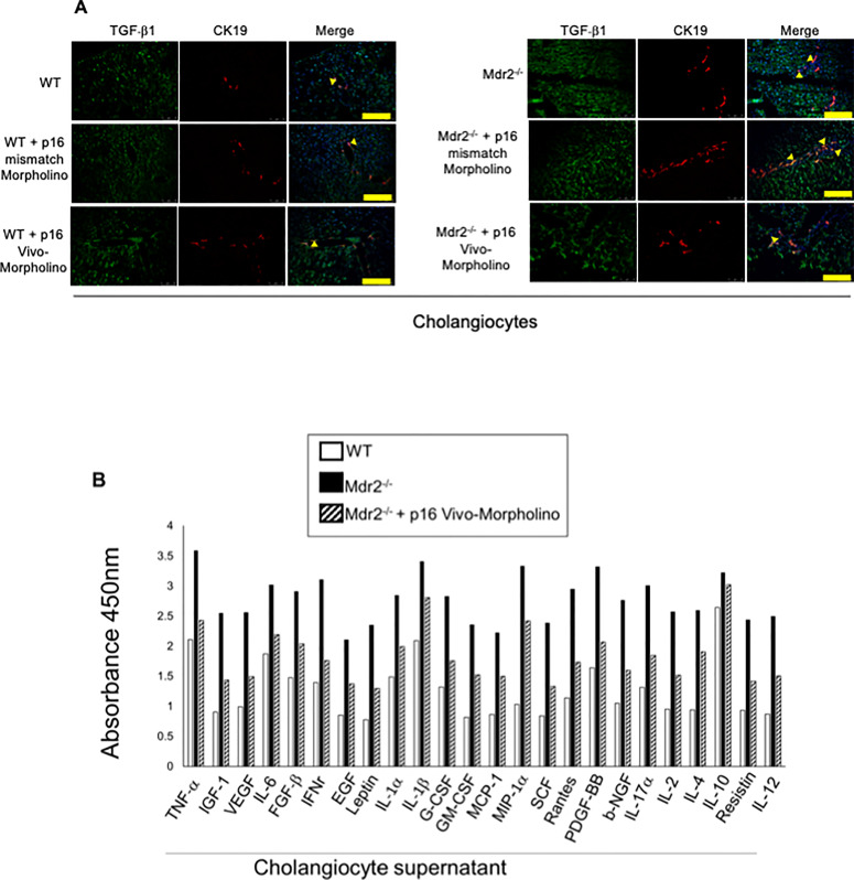
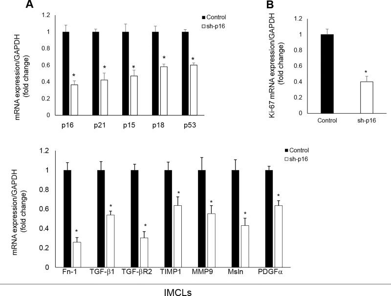
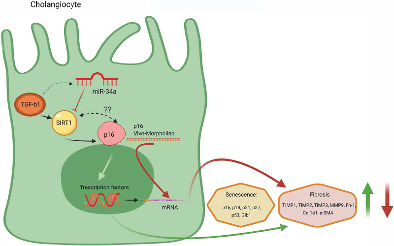
Biliary senescence and hepatic fibrosis are hallmarks of cholangiopathies including primary sclerosing cholangitis (PSC). Senescent cholangiocytes display senescence-associated secretory phenotypes [SASPs, e.g., transforming growth factor-1 (TGF-1)] that further increase biliary senescence (by an autocrine loop) and trigger liver fibrosis by paracrine mechanisms. The aim of this study was to determine the effect of p16 inhibition and role of the TGF-1/microRNA (miR)-34a/sirtuin 1 (SIRT1) axis in biliary damage and liver fibrosis in the Mdr2/ mouse model of PSC. We treated (i) in vivo male wild-type (WT) and Mdr2/ mice with p16 Vivo-Morpholino or controls before measuring biliary mass [intrahepatic bile duct mass (IBDM)] and senescence, biliary SASP levels, and liver fibrosis, and (ii) in vitro intrahepatic murine cholangiocyte lines (IMCLs) with small interfering RNA against p16 before measuring the mRNA expression of proliferation, senescence, and fibrosis markers. p16 and miR-34a increased but SIRT1 decreased in Mdr2/ mice and PSC human liver samples compared to controls. p16 immunoreactivity and biliary senescence and SASP levels increased in Mdr2/ mice but decreased in Mdr2/ mice treated with p16 Vivo-Morpholino. The increase in IBDM and hepatic fibrosis (observed in Mdr2/ mice) returned to normal values in Mdr2/ mice treated with p16 Vivo-Morpholino. TGF-1 immunoreactivity and biliary SASPs levels were higher in Mdr2/ compared to those of WT mice but returned to normal values in Mdr2/ mice treated with p16 Vivo-Morpholino. The expression of fibrosis/senescence markers decreased in cholangiocytes from Mdr2/ mice treated with p16 Vivo-Morpholino (compared to Mdr2/ mice) and in IMCLs (after p16 silencing) compared to controls. Modulation of the TGF-1/miR-34a/SIRT1 axis may be important in the management of PSC phenotypes.


Figures

Figure 1
Figure 1
Representative diagram of the microRNA (miR)-34a/sirtuin 1 (SIRT1)/p16 axis outlined using the IPA pathway. The IPA-generated IPA supports our hypothesis showing that miR-34a modulates p16 (marker of senescence) through changes in SIRT1. Reproduced with permission from Qiagen.
Figure 2
Figure 2
By immunofluorescence, we demonstrated increased immunoreactivity for p16 in bile ducts of Mdr2−/− mice and Mdr2−/− mice treated with p16 mismatch Morpholino compared to their respectively wild-type (WT) and Mdr2−/−-treated mismatch Morpholino; p16 (red staining); CK19 (green staining); localization of p16 in bile ducts is indicated by yellow arrows. Nuclei were stained with 4′,6-diamidino-2-phenylindole (DAPI). Scale bar: 100 μm.
Figure 3
Figure 3
(A) There was enhanced p16 biliary immunoreactivity in Mdr2−/− compared to normal WT mice, which was decreased in Mdr2−/− mice treated with p16 mismatch Vivo-Morpholino compared to Mdr2−/− mice treated with mismatch Morpholino. Original magnification: 20×. (B) By SA-β-galactosidase (SA-β-GAL) staining in liver sections, there was enhanced biliary senescence in Mdr2−/− compared to WT mice that decreased in Mdr2−/− mice treated with p16 Vivo-Morpholino compared to mismatch-treated Mdr2−/− mice. Original magnification: 10×. (A, B) No changes in p16 immunoreactivity and biliary senescence immunoreactivity were observed between WT mice treated with p16 Vivo-Morpholino and mismatch control. (C) By real-time polymerase chain reaction (PCR), the mRNA expression of p15, p18, p21, p27, p53, and Glb1 increased in cholangiocytes from Mdr2−/− compared to WT mice, but decreased in Mdr2−/− mice treated with p16 Vivo-Morpholino compared to Mdr2−/− mice. Data are mean ± standard error of the mean (SEM) of three evaluations performed in a cumulative preparation of cholangiocytes from four mice *p < 0.05 versus WT mice. #p < 0.05 versus Mdr2−/− mice.
Figure 4
Figure 4
IBDM increased in Mdr2−/− compared to WT mice, but decreased in Mdr2−/− mice treated with p16 Vivo-Morpholino mice compared to their respective mismatched controls. In Mdr2−/− mice, IBDM increased significantly compared to WT mice; however, the administration of p16 Vivo-Morpholino markedly decreased IBDM compared to Mdr2−/− mice treated with mismatch Morpholino; IBDM was similar among WT mice treated with p16 Vivo-Morpholino or the respective mismatch control. Bile ducts are indicated by red arrows. Ten different fields from three samples from three different animals were analyzed. Original magnification: 20×. *p < 0.05 versus WT mice. #p < 0.05 versus Mdr2−/− mice.
Figure 5
Figure 5
(A) There was increased collagen deposition in Mdr2−/− compared to WT mice, which decreased in Mdr2−/− mice treated with p16 Vivo-Morpholino compared to Mdr2−/− mice treated with mismatch Morpholino. Ten different fields from three samples from three different animals were analyzed. Original magnification: 20×. *p < 0.05 versus WT mice. #p < 0.05 versus Mdr2−/− mice. Black arrows show collagen deposition around bile ducts. (B) By immunofluorescence, there was increased immunoreactivity of Col1a1 in Mdr2−/− compared to WT mice, which decreased in Mdr2−/− mice treated with p16 Vivo-Morpholino sequences compared to Mdr2−/− mice treated with mismatch Morpholino. Col1a1 (green staining); CK19 (red staining); nuclei were stained with DAPI; scale bar: 100 μm. *p < 0.05 versus WT mice. #p < 0.05 versus Mdr2−/− mice. (A, B). There were no changes in liver fibrosis between WT mice treated with p16 mismatch Morpholino and p16 Vivo-Morpholino. (C) By real-time PCR, the mRNA expression of fibrosis markers Fn-1, Col1a1, a-SMA, TIMP1, TIMP2, TIMP3, and MMP9 increased in cholangiocytes from Mdr2−/− compared to WT mice, but decreased in Mdr2−/− mice treated with p16 Vivo-Morpholino compared to Mdr2−/− mice. Data are mean ± SEM of three evaluations performed in a cumulative preparation of cholangiocytes from four mice. *p < 0.05 versus WT mice. #p < 0.05 versus Mdr2−/− mice.
Figure 6
Figure 6
(A, B) In human PSC samples, we demonstrated (i) enhanced expression of miR-34a (in total liver samples) and (ii) reduced mRNA expression of SIRT1 in total liver samples compared to normal human samples. Data are mean ± SEM from seven late stage PSC patients and five normal human controls. *p < 0.05 versus control human samples. (C) By real-time PCR, there was reduced levels of SIRT1 in isolated cholangiocytes and total liver from Mdr2−/− mice compared to WT mice, which returned to values similar to those of WT mice in Mdr2−/− mice treated with p16 Vivo-Morpholino. Data are mean ± SEM of three evaluations performed in a cumulative preparation of cholangiocytes from four mice. Data are mean ± SEM of three evaluations performed in three different total liver samples from three different mice. *p < 0.05 versus WT mice, #p < 0.05 versus Mdr2−/− mice.
Figure 7
Figure 7
(A) By immunofluorescence, we demonstrated increased immunoreactivity for transforming growth factor-β1 (TGF-β1) in bile ducts of Mdr2−/− mice and Mdr2−/− mice treated with p16 mismatch Morpholino compared to their respective WT and Mdr2−/− treated mismatch Morpholino; TGF-β1 (green staining); CK19 (red staining); localization of TGF-β1 in bile ducts is indicated by yellow arrows. Nuclei were stained with DAPI. Scale bar: 100 μm. (B) There was increase in SASPs levels in cholangiocyte supernatant from Mdr2−/− mice (compared to WT mice), parameters that all decreased in Mdr2−/− mice treated with p16 Vivo-Morpholino compared to Mdr2−/− mice. Evaluation is the result of cumulative preparation of cholangiocytes from four mice.
Figure 8
Figure 8
(A, B) In p16-silenced IMCLs, there was reduced expression of senescence, fibrosis, and proliferation (Ki-67) mRNAs compared to control IMCLs. Data are mean ± SEM of n = 3 from three preparations of IMCLs. *p < 0.05 versus IMCLs.
Figure 9
Figure 9
We depicted a working model illustrating the role of the TGF-β1/miR-34a/SIRT1/p16 axis in regulating biliary proliferation/senescence and paracrine activation of hepatic stellate cells (HSCs) with subsequent increase in liver fibrosis. TGF-β1 stimulates miR-34a and inhibits SIRT1, activating p16 that stimulates the transcription of the senescence (p15, p18, p21, p27, p53, and Glb1) and fibrosis (TIMP1, TIMP2, MMP9, Fn-1, Col1a1, and α-SMA) markers. Also, TGF-β1 stimulates directly p16 and activates the fibrosis and senescence gene transcription. The cartoon suggests a link between TGF-β1, p16, and SIRT1 through a feedback mechanism. Created with BioRender. com.

Similar articles

Cited by

References

    1. Alpini G, Lenzi R, Sarkozi L, Tavoloni N. Biliary physiology in rats with bile ductular cell hyperplasia. Evidence for a secretory function of proliferated bile ductules. J Clin Invest. 1988;81(2):569–78. - PMC - PubMed
    1. Kanno N, LeSage G, Glaser S, Alpini G. Regulation of cholangiocyte bicarbonate secretion. Am J Physiol Gastrointest Liver Physiol. 2001;281(3):G612–25. - PubMed
    1. Glaser S, Gaudio E, Miller T, Alvaro D, Alpini G. Cholangiocyte proliferation and liver fibrosis. Expert Rev Mol Med. 2009;11:e7. - PMC - PubMed
    1. Tabibian JH, O’Hara SP, Splinter PL, Trussoni CE, LaRusso NF. Cholangiocyte senescence by way of N-ras activation is a characteristic of primary sclerosing cholangitis. Hepatology 2014;59(6):2263–75. - PMC - PubMed
    1. Nakanuma Y, Sasaki M, Harada K. Autophagy and senescence in fibrosing cholangiopathies. J Hepatol. 2015;62(4):934–45. - PubMed

Publication types

MeSH terms