. 2020 Jul 10;295(28):9349-9365.
doi: 10.1074/jbc.RA120.013630. Epub 2020 May 11.

Retargeting from the CR3 to the LFA-1 receptor uncovers the adenylyl cyclase enzyme-translocating segment of Bordetella adenylate cyclase toxin

Affiliations

Retargeting from the CR3 to the LFA-1 receptor uncovers the adenylyl cyclase enzyme-translocating segment of Bordetella adenylate cyclase toxin

Jiri Masin et al. J Biol Chem. .

Abstract

The Bordetella adenylate cyclase toxin-hemolysin (CyaA) and the α-hemolysin (HlyA) of Escherichia coli belong to the family of cytolytic pore-forming Repeats in ToXin (RTX) cytotoxins. HlyA preferentially binds the αLβ2 integrin LFA-1 (CD11a/CD18) of leukocytes and can promiscuously bind and also permeabilize many other cells. CyaA bears an N-terminal adenylyl cyclase (AC) domain linked to a pore-forming RTX cytolysin (Hly) moiety, binds the complement receptor 3 (CR3, αMβ2, CD11b/CD18, or Mac-1) of myeloid phagocytes, penetrates their plasma membrane, and delivers the AC enzyme into the cytosol. We constructed a set of CyaA/HlyA chimeras and show that the CyaC-acylated segment and the CR3-binding RTX domain of CyaA can be functionally replaced by the HlyC-acylated segment and the much shorter RTX domain of HlyA. Instead of binding CR3, a CyaA1-710/HlyA411-1024 chimera bound the LFA-1 receptor and effectively delivered AC into Jurkat T cells. At high chimera concentrations (25 nm), the interaction with LFA-1 was not required for CyaA1-710/HlyA411-1024 binding to CHO cells. However, interaction with the LFA-1 receptor strongly enhanced the specific capacity of the bound CyaA1-710/HlyA411-1024 chimera to penetrate cells and deliver the AC enzyme into their cytosol. Hence, interaction of the acylated segment and/or the RTX domain of HlyA with LFA-1 promoted a productive membrane interaction of the chimera. These results help delimit residues 400-710 of CyaA as an "AC translocon" sufficient for translocation of the AC polypeptide across the plasma membrane of target cells.

Keywords: AC domain translocation; AC translocon; Bordetella pertussis; CyaA; Escherichia coli (E. coli); HlyA; RTX toxin; acylation; acyltransferase; bacterial toxin; complement receptor 3 (CR3,); fatty acid; fatty acyl; integrin; protein acylation; protein translocation.


Conflict of interest statement

Conflict of interest—The authors declare no conflicts of interest in regards to this manuscript.

Figures

Figure 1.
Figure 1.
Schematic representation of CyaA, HlyA, and hybrid CyaA/HlyA molecules. Individual domains of CyaA and HlyA are indicated by the colored rectangles. AC, adenylate cyclase domain; LS, AC-to-Hly linker segment; PF, pore-forming domain; AS, acylated segment; RTX, calcium-binding repeats; SS, secretion signal. The numbers that follow the CyaA or HlyA in the names of the CyaA/HlyA hybrid chimera represent the number of the first and last residue of the segment of the given protein according to the sequences of full-length CyaA and HlyA, respectively. Design of the hybrid molecules: (i) in CyaA1-501/HlyA142-1024, the AC domain and the adjacent AC-to-Hly linker segment of CyaA (residues 1-501 of CyaA) are fused to a sequence beginning with the first putative transmembrane α-helix of HlyA (residues 142-1024 of HlyA); (ii) in CyaA1-528/HlyA238-1024, the AC domain, the adjacent AC-to-Hly linker segment, and the first α-helix of CyaA (residues 1-528 of CyaA) are fused to a sequence beginning with the second putative α-helix of HlyA (residues 238-1024 of HlyA); (iii) in CyaA1-606/HlyA368-1024, the AC domain, the adjacent AC-to-Hly linker segment, and the first three α-helices of CyaA (residues 1-606 of CyaA) are fused to a sequence beginning with the fourth putative α-helix of HlyA (residues 368-1024 of HlyA); (iv) in CyaA1-710/HlyA411-1024, the AC domain, the adjacent AC-to-Hly linker segment and the hydrophobic domain of CyaA (1-710 of CyaA) are fused to a sequence beginning with the acylated segment of HlyA (residues 411-1024 of HlyA); (v) in CyaA1-800/HlyA501-1024, the AC domain, the adjacent AC-to-Hly linker segment, the hydrophobic domain, and part of the acylated segment of CyaA (residues 1- 800 of CyaA) are fused to a sequence beginning with the truncated acylated segment of HlyA (residues 501-1024 of HlyA).
Figure 2.
Figure 2.
16-Carbon mono- and 14-carbon doubly-acylated (CyaC- and HlyC-activated) hybrid molecules bind and lyse erythrocytes with importantly differing efficacies. A, sheep erythrocytes (5 × 10⁸/ml) were incubated in the presence of 75 mm sucrose as osmoprotectant with 5 nm purified proteins at 37 °C and after 30 min, aliquots were taken for determinations of the cell-associated AC activity (Binding). Activities are expressed as percentages of the activity of the intact 16-carbon doubly-acylated, CyaC-activated CyaA and represent average mean ± S.D. from at least three independent determinations performed in duplicate with two different toxin preparations. Sheep erythrocytes (5 × 10⁸/ml) were incubated at 37 °C in the presence of CyaC-activated (B) or HlyC-activated (C) proteins (25 nm). Hemolytic activity was measured as the amount of released hemoglobin by photometric determination (A541) (n = 3).
Figure 3.
Figure 3.
Nonhemolytic HlyC-activated chimeras CyaA1-710/HlyA411-1024, and CyaA1-800/HlyA501-1024 translocate the AC domain across the plasma membrane of sheep erythrocytes in a calcium-dependent manner. A, sheep erythrocytes (5 × 10⁸/ml) were incubated in the presence of 75 mm sucrose as osmoprotectant with 5 nm purified proteins at 37 °C and after 30 min, aliquots were taken for determinations of the AC activity internalized into erythrocytes and protected against digestion by externally added trypsin (Invasive AC). Activities are expressed as the activity of the intact, 16-carbon doubly-acylated, CyaC-activated CyaA and represent average mean ± S.D. from at least three independent determinations performed in duplicate with two different toxin preparations. B, cAMP intoxication was assessed by determining the intracellular concentration of cAMP generated in sheep erythrocytes (5 × 10⁸/ml) after 30 min of incubation of cells with 25 nm CyaA or the hybrid proteins. Activities are expressed as percentages of intact CyaC-activated CyaA activity and represent average mean ± S.D. from four independent determinations performed in duplicate. C, sheep erythrocytes (5 × 10⁸/ml) were incubated in the presence of 2 mm calcium (+Ca2+) or in absence of calcium and presence of 5 mm EDTA (+EDTA) at 37 °C with 5 nm proteins. After 30 min, aliquots were taken for determinations of the cell-associated AC activity (Binding) and AC activity internalized into erythrocytes and protected against digestion by externally added trypsin (Invasive AC). Activities are expressed as percentages of intact CyaC-activated CyaA activity (n = 3-7).
Figure 4.
Figure 4.
The cell-invasive CyaA1-710/HlyA411-1024 hybrid molecules exhibit a very low membrane-permeabilizing activity. A, overall membrane activities of intact CyaC-acylated CyaA and CyaC- or HlyC-activated CyaA1-710/HlyA411-1024 hybrid molecules on asolectin/decane:butanol (9:1) membranes. Conditions of measurement were: 150 mm KCl, 10 mm Tris-HCl (pH 7.4), 2 mm CaCl2; the applied voltage was 50 mV; temperature was 25 °C, and the recording was filtered at 10 Hz; the protein concentration was 250 pm. B, single-pore recordings of asolectin membranes in the presence of 10 pm purified protein variants under otherwise identical conditions as in A.
Figure 5.
Figure 5.
The CyaA1-710/HlyA411-1024 hybrid preferentially binds LFA-1–positive cells and monoacylation by CyaC is sufficient for AC translocating activity. A, binding of the intact CyaC-activated CyaA or HlyC-activated CyaA1-710/HlyA411-1024 hybrid variant to J774A.1 mouse macrophages (1 × 10⁶) was determined as the amount of total cell-associated AC enzyme activity upon incubation of cells with 5 nm protein for 30 min at 4 °C. To block the binding site of CyaA on the CR3 receptor, the J774A.1 cells (1 × 10⁶) were preincubated for 30 min on ice with 5 μg/ml of the CD11b-specific mAb M1/70 prior to addition of 5 nm CyaA (CyaC+) or CyaA1-710/HlyA411-1024 (HlyC+). Activities are expressed as percentages of intact CyaA activity in the absence of M1/70 mAb and represent average mean ± S.D. from two independent determinations performed in duplicate with two different toxin preparations. B, cAMP intoxication was assessed by determining the intracellular concentration of cAMP after 30 min of incubation of J774A.1 cells (2 × 10⁵) with toxins (n = 3). C, binding of CyaA or CyaA1-710/HlyA411-1024 (HlyC+) to Jurkat T-cells (1 × 10⁶) was determined as the amount of total cell-associated AC enzyme activity upon incubation of cells with 5 and 50 nm toxin for 30 min at 4 °C. Activities are expressed as percentages of intact CyaA activity and represent average mean ± S.D. from two independent determinations performed in duplicate with two different toxin preparations. D, cAMP intoxication was assessed by determining the intracellular concentration of cAMP generated in Jurkat T-cells (3 × 10⁵) after 30 min of incubation at different toxin concentrations (n = 5). E, binding of CyaA1-710/HlyA411-1024 (HlyC+) or intact CyaA to CHO cells expressing CD11a/CD18 or mock-transfected CHO cells (1 × 10⁶) was determined as the amount of total cell-associated AC enzyme activity upon incubation of cells with indicated toxin concentrations for 30 min at 4 °C. Activities represent average mean ± S.D. from three independent determinations performed in duplicate with two different toxin preparations. F, cAMP intoxication was assessed by determining the intracellular concentration of cAMP generated in CHO cells expressing CD11a/CD18 or mock-transfected CHO cells after 30 min of incubation of cells (1 × 10⁵) with different concentrations of CyaA1-710/HlyA411-1024 (HlyC+) or intact CyaA (n = 4). *, statistically significant differences (p < 0.05); **, statistically significant differences (p < 0.01); ***, statistically significant differences (p < 0.001); ****, statistically significant differences (p < 0.0001).
Figure 6.
Figure 6.
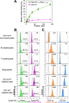
Binding of fluorescently labeled CyaA-AC1-710/HlyA411-1024 and CyaA-AC to spleen cells in vitro. A, spleen single cell suspensions (1 × 10⁶) from 7-week–old Balb/c mice were incubated in vitro with a range of equimolar concentrations of separately added Dy495-labeled CyaA-AC1-710/HlyA411-1024 or Dy650-labeled CyaA-AC and were analyzed by flow cytometry (for gating strategy see Fig. S5). B, spleen single cell suspensions (1 × 10⁶) from 7-week-old Balb/c mice were incubated with an equimolar mixture of 56 nm fluorescently-labeled or nonfluorescent CyaA-AC1-710/HlyA411-1024 and CyaA-AC for 30 min at 4 °C. Cells were then stained with a mixture of fluorescently-labeled antibodies against cell-surface antigens. Binding of CyaA-AC1-710/HlyA411-1024–Dy495 or CyaA-AC–Dy650 to nonleukocytic cells (CD11a), B lymphocytes (CD19+), T lymphocytes (CD3+), neutrophils (Ly-6G+CD11b+), CD11bhigh myeloid cells (Lineageneg CD11bhigh) and CD11bint myeloid cells (Lineageneg CD11bint) was determined by flow cytometry and depicted as histograms of Dy650 or Dy495 signal in each population. Gray histograms represent nonfluorescent CyaA-AC1-710/HlyA411-1024 or CyaA-AC controls. Dy650- or Dy495-positive cells in each population are shown in percentages. Values in top right corners indicate the mean fluorescence intensity (MFI) for each histogram with subtracted MFI of nonfluorescent controls. C, spleen single cell suspensions from 7-week-old Balb/c mice were stained with a mixture of fluorescently labeled antibodies against cell-surface antigens. Expression of CD11a and CD11b on corresponding cell populations was determined by flow cytometry. Gray histograms represent corresponding isotype control. Values in top right corners indicate mean fluorescent intensity for each histogram with subtracted mean fluorescent intensity of isotype controls. A result from one representative experiment is shown in each panel. In vitro experiment was repeated two times independently with similar results.
Figure 7.
Figure 7.
Schematic representation of CyaA and HlyA domains sufficient for AC translocation and LFA-1 binding. A, the AC domain of Bordetella pertussis CyaA has to be fused to the adjacent AC-to-Hly linker segment and to the five predicted transmembrane α-helices for an efficient translocation across the cell membrane, whereas the acylated segment and the RTX domain segment of CyaA can be replaced with the corresponding segments of the E. coli HlyA toxin. B, the doubly acylated HlyC-modified 14-carbon-acylated CyaA1-710/HlyA411-1024 hybrid, bearing C14:0 or C14:0-OH on the two Lys-564 and Lys-690 residues translocated the AC domain into cell cytosol of both, LFA-1–positive and LFA-1–negative cells. C, the monoacylated CyaA1-710/HlyA411-1024 hybrid, bearing C16:0, C16:1, and/or C18:1 on the Lys-690 residue intoxicated the LFA-1–positive cells with the same efficacy as the double acylated hybrid but was poorly active on erythrocytes lacking the β2 integrin LFA-1. Because CyaA does not bind the LFA-1 integrin (Fig. S3), the LFA-1–binding segment of the HlyC-acylated CyaA1-710/HlyA411-1024 molecule is confined in the acylated segment and/or the RTX domain of HlyA (residues 411 to 1024).

References

    1. Linhartová I., Bumba L., Mašín J., Basler M., Osička R., Kamanova J., Prochazkova K., Adkins I., Hejnová-Holubová J., Sadilková L., Morová J., and Sebo P. (2010) RTX proteins: a highly diverse family secreted by a common mechanism. FEMS Microbiol. Rev. 34, 1076–1112 10.1111/j.1574-6976.2010.00231.x - DOI - PMC - PubMed
    1. Novak J., Cerny O., Osickova A., Linhartova I., Masin J., Bumba L., Sebo P., and Osicka R. (2017) Structure-function relationships underlying the capacity of Bordetella adenylate cyclase toxin to disarm host phagocytes. Toxins 9, 300 10.3390/toxins9100300 - DOI - PMC - PubMed
    1. Sebo P., Osicka R., and Masin J. (2014) Adenylate cyclase toxin-hemolysin relevance for pertussis vaccines. Exp. Rev. Vaccines 13, 1215–1227 10.1586/14760584.2014.944900 - DOI - PubMed
    1. Vojtova J., Kamanova J., and Sebo P. (2006) Bordetella adenylate cyclase toxin: a swift saboteur of host defense. Curr. Opin. Microbiol. 9, 69–75 10.1016/j.mib.2005.12.011 - DOI - PubMed
    1. Gordon V. M., Leppla S. H., and Hewlett E. L. (1988) Inhibitors of receptor-mediated endocytosis block the entry of Bacillus anthracis adenylate cyclase toxin but not that of Bordetella pertussis adenylate cyclase toxin. Infect. Immun. 56, 1066–1069 10.1128/IAI.56.5.1066-1069.1988 - DOI - PMC - PubMed

Publication types

Substances