. 2020 May 26;117(21):11667-11673.
doi: 10.1073/pnas.1911870117. Epub 2020 May 11.

Hepatic gap junctions amplify alcohol liver injury by propagating cGAS-mediated IRF3 activation

Affiliations

Hepatic gap junctions amplify alcohol liver injury by propagating cGAS-mediated IRF3 activation

Jay Luther et al. Proc Natl Acad Sci U S A. .

Erratum in

Abstract

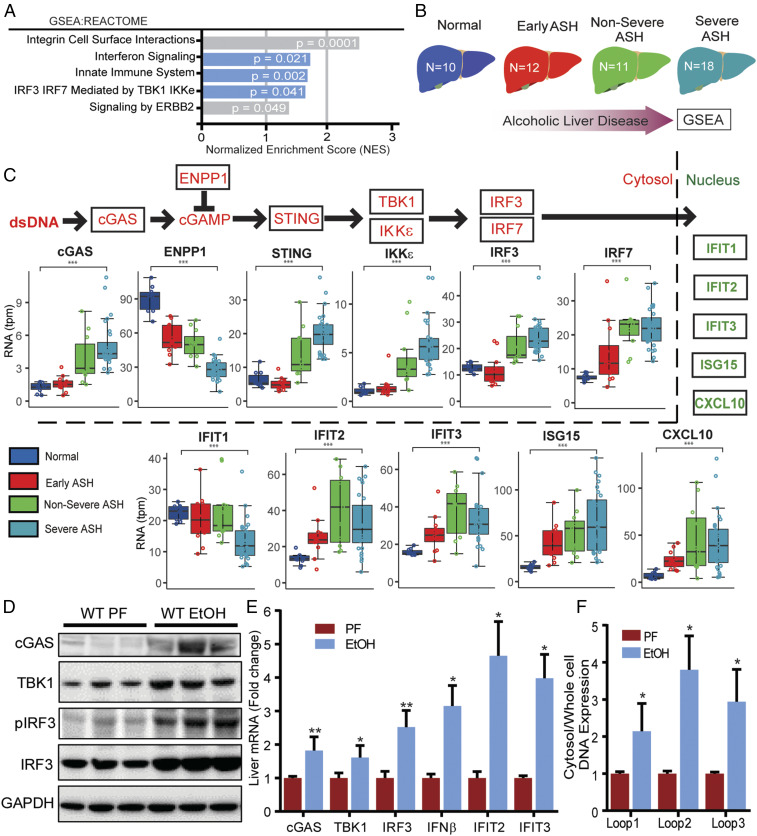
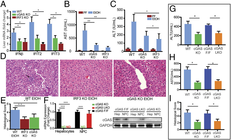
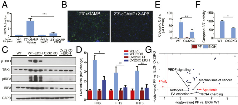
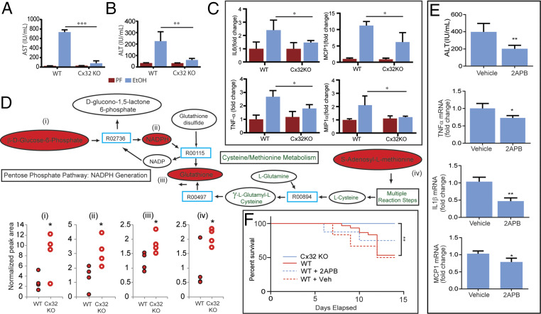
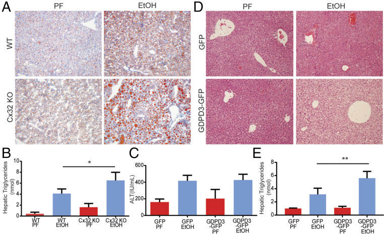
Alcohol-related liver disease (ALD) accounts for the majority of cirrhosis and liver-related deaths worldwide. Activation of IFN-regulatory factor (IRF3) initiates alcohol-induced hepatocyte apoptosis, which fuels a robust secondary inflammatory response that drives ALD. The dominant molecular mechanism by which alcohol activates IRF3 and the pathways that amplify inflammatory signals in ALD remains unknown. Here we show that cytoplasmic sensor cyclic guanosine monophosphate-adenosine monophosphate (AMP) synthase (cGAS) drives IRF3 activation in both alcohol-injured hepatocytes and the neighboring parenchyma via a gap junction intercellular communication pathway. Hepatic RNA-seq analysis of patients with a wide spectrum of ALD revealed that expression of the cGAS-IRF3 pathway correlated positively with disease severity. Alcohol-fed mice demonstrated increased hepatic expression of the cGAS-IRF3 pathway. Mice genetically deficient in cGAS and IRF3 were protected against ALD. Ablation of cGAS in hepatocytes only phenocopied this hepatoprotection, highlighting the critical role of hepatocytes in fueling the cGAS-IRF3 response to alcohol. We identified connexin 32 (Cx32), the predominant hepatic gap junction, as a critical regulator of spreading cGAS-driven IRF3 activation through the liver parenchyma. Disruption of Cx32 in ALD impaired IRF3-stimulated gene expression, resulting in decreased hepatic injury despite an increase in hepatic steatosis. Taken together, these results identify cGAS and Cx32 as key factors in ALD pathogenesis and as potential therapeutic targets for hepatoprotection.

Keywords: IRF3; alcohol liver; cGAS; connexin; innate immunity.


Conflict of interest statement

Competing interest statement: The authors declare a competing interest. K.R.K., M.Y.L., and S.J.P. have equity interest in Heprotech Inc. M.K.G. has equity interest in New Amsterdam Genomics. All other authors have no conflicts to declare.

Figures

Fig. 1.
Fig. 1.
Alcohol activates the cGAS-IRF3 pathway in humans and mice. (A) GSEA of RNA-seq expression data from liver tissue of healthy controls and patients with alcohol-related liver disease (ALD). The enrichment score (ES) reflects the degree to which a gene set is overrepresented at the top or bottom of a ranked list of genes. (B) Schematic depicting the spectrum of disease severity within the ALD patient cohort. (C, Top) Abbreviated schematic of the cGAS-IRF3 pathway (red) and downstream IRF3-regulated genes (green). (C, Bottom) Differential expression analysis of cGAS-IRF3 pathway and IRF3-regulated genes based on disease severity within the ALD patient cohort. WT mice were fed 5% alcohol (EtOH) or a control PF diet for 6 wk. (D) Immunoblot for cGAS, TBK1, pIRF3, IRF3, and GAPDH and (E) hepatic mRNA expression of cGas, Tbk1, Irf3, and IRF3-regulated genes (Ifnβ, Ifit2, Ifit3) from EtOH and PF WT liver tissue. DNA in the cytosolic fraction hepatocytes from WT ETOH- and PF-fed mice was isolated, and the relative abundance of mtDNA was measured by RT-PCR normalized to whole-cell extract. (F) Cytosolic mtDNA content in liver tissue from WT and PF mice. N = 10 mice/group. Data are shown as mean ± SD. *P < 0.05; **P < 0.01; ***P < 0.001.
Fig. 2.
Fig. 2.
Hepatocyte-specific cGAS drives alcohol-induced IRF3 activation and liver injury. WT, cGAS-deficient (cGAS KO), and IRF3-deficient (IRF3 KO) mice were exposed to the 10-d NIAAA chronic and binge alcohol model. (A) Hepatic mRNA expression of IRF3-regulated genes (Ifnβ, Ifit2, Ifit3), (B and C) serum transaminase levels, and (D and E) liver histology and histological scoring (hematoxylin and eosin [H&E], 40× magnification; arrow highlighting an inflammatory foci) from WT, cGAS KO, and IRF3 KO mice. (F) mRNA expression and immunoblot for cGas in hepatocytes and nonparenchymal cells (NPCs) from cGAS F/F, cGAS KO, and cGAS LKO mice. (G and H) Serum transaminase levels and (I) histological score from WT, cGAS KO, cGAS F/F, and cGAS LKO mice exposed to the NIAAA chronic and binge alcohol model. N = 6–10 mice/group. Data are shown as mean ± SD. *P < 0.05; **P < 0.01; ***P < 0.001.
Fig. 3.
Fig. 3.
Cx32 amplifies alcohol-induced IRF3 activation and apoptosis. Hepatocyte-derived H35 cells were stimulated with 2′3′-cGAMP or vehicle (donor cells) and cocultured with unstimulated or 2APB–treated H35 IRF3-GFP reporter cells (recipient cells) that express GFP upon IRF3 activation. (A) IRF3 activity as determined by flow cytometry and (B) representative 20× fluorescence images of 2′3′-cGAMP–stimulated H35 cells cocultured with H35 GFP reporter cells in the presence or absence of 2APB (green = GFP, blue = DAPI). Cx32KOmice and their littermate controls (WT) were exposed to the 10-d NIAAA chronic and binge alcohol model. (C) Immunoblot for phosphorylated TBK1 (pTBK1), TBK1, pIRF3, IRF3, and GAPDH, (D) hepatic mRNA expression of IRF3-regulated genes (Ifnβ, Ifit2, Ifit3), (E) cytosolic cytochrome C activity, and (F) Caspase 3/7 in liver tissue from WT and Cx32KO mice. (G) Ingenuity pathway analysis (IPA) was used to analyze hepatic transcriptome data for the following two comparisons: 1) ethanol vs. sober for WT mice and 2) ethanol vs. sober for Cx32KO mice. PEA provided P values associated with a pathway based on Fisher’s exact test and plotted in negative log scale for both comparisons. N = 10 mice/group. Data are shown as mean ± SD. *P < 0.05; **P < 0.01; ***P < 0.001.
Fig. 4.
Fig. 4.
Cx32 propagates alcohol-induced hepatocyte injury, inflammation, and mortality. WT and Cx32KO mice were exposed to the 10-d NIAAA chronic and binge ethanol model. (A and B) Serum transaminase levels and (C) hepatic mRNA expression of inflammatory genes (Il6, Mcp1, Tnfα, Mip1α) from WT and Cx32KO mice. (D) Hepatic metabolomic profiling of key mediators involved in oxidative stress pathways in WT and Cx32KO mice. (E) WT mice were exposed to the 10-d NIAAA chronic and binge ethanol model and treated with an intraperitoneal injection of 2APB (20 mg/kg) or vehicle control (dimethyl sulfoxide [DMSO], 0.1 mL/kg) prior to oral gavage of ethanol on day 10. Serum ALT and hepatic mRNA expression of inflammatory genes (Il1β, Mcp1, Tfnα). (F) Kaplan-Meier survival curve for 1) Cx32KO mice, 2) WT mice, 3) WT mice treated with daily 2APB injections (20 mg/kg), and 4) WT mice treated with daily vehicle injections of DMSO (0.1 mL/kg), exposed to a 6% ethanol diet. N = 6–10 mice/group. Data are shown as mean ± SD. *P < 0.05; **P < 0.01; ***P < 0.001.
Fig. 5.
Fig. 5.
Cx32 deficiency induces hepatic triglyceride accumulation and up-regulation of GDPD3. (A and B) WT and Cx32-deficient (Cx32KO) mice were exposed to the chronic and binge ethanol model. (A) Histological evidence of increased lipid deposition in Cx32KO mice compared to WT mice based on Oil Red O staining. (B) Increased hepatic triglyceride content in Cx32KO mice compared to WT mice. (CE) WT mice were exposed to a modified version of the chronic and binge ethanol model, in which mice were injected on day 9 with either recombinant adenoviral vectors expressing GDPD3-GFP or GFP alone. Mice were then gavaged with 5 g ethanol/kg body weight on day 12 and killed 9 h later. (C) No difference in serum ALT in GDPD3-GFP–infected mice compared to control GFP-infected mice. (D) Increased steatosis based on histological analysis of H&E specimens. (E) Levels of hepatic triglycerides in GDPD3-GFP–infected mice compared to control GFP-infected mice. (Data are shown as mean ± SD and analyzed by Student’s t tests. N = 8–12 mice/group. *P < 0.05; **P < 0.01; ***P < 0.001.

References

    1. Schwartz J. M., Reinus J. F., Prevalence and natural history of alcoholic liver disease. Clin. Liver Dis. 16, 659–666 (2012). - PubMed
    1. Nguyen-Khac E., et al. ; AAH-NAC Study Group , Glucocorticoids plus N-acetylcysteine in severe alcoholic hepatitis. N. Engl. J. Med. 365, 1781–1789 (2011). - PubMed
    1. Gao B., et al. , Innate immunity in alcoholic liver disease. Am. J. Physiol. Gastrointest. Liver Physiol. 300, G516–G525 (2011). - PMC - PubMed
    1. Nagy L. E., The role of innate immunity in alcoholic liver disease. Alcohol Res. 37, 237–250 (2015). - PMC - PubMed
    1. Szabo G., Petrasek J., Bala S., Innate immunity and alcoholic liver disease. Dig. Dis. 30 (suppl. 1), 55–60 (2012). - PMC - PubMed

MeSH terms

Substances