. 2020 May 26;117(21):11788-11798.
doi: 10.1073/pnas.1921314117. Epub 2020 May 11.

Prefrontal-hippocampal functional connectivity encodes recognition memory and is impaired in intellectual disability

Affiliations

Prefrontal-hippocampal functional connectivity encodes recognition memory and is impaired in intellectual disability

Maria Alemany-González et al. Proc Natl Acad Sci U S A. .

Abstract

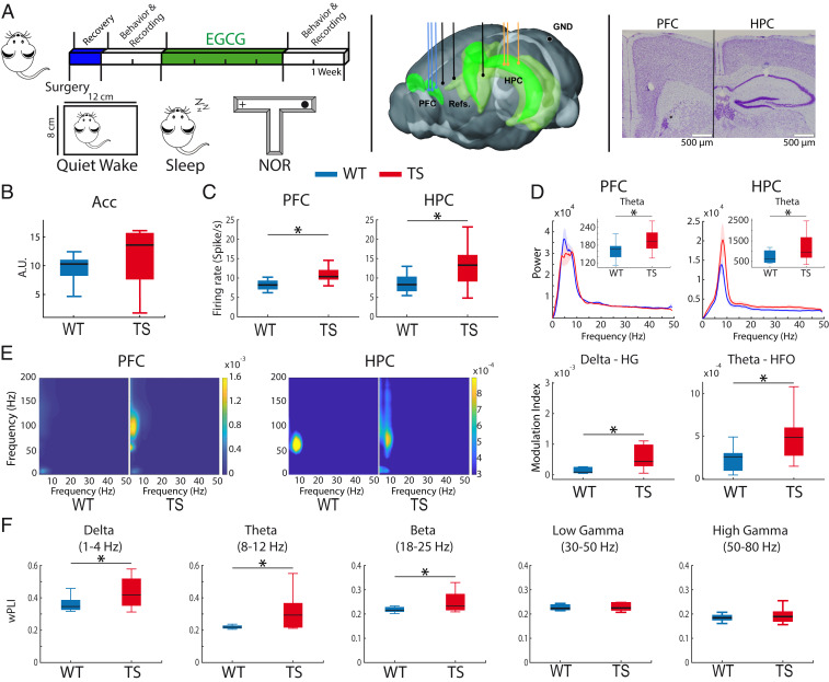
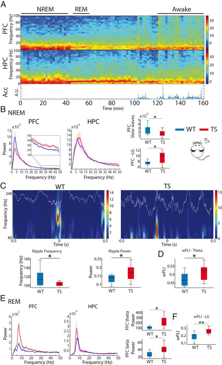
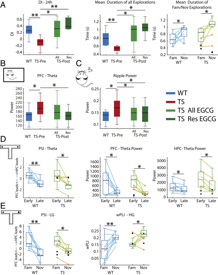
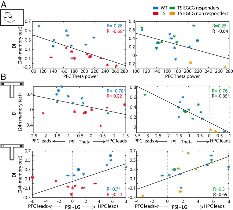
Down syndrome (DS) is the most common form of intellectual disability. The cognitive alterations in DS are thought to depend on brain regions critical for learning and memory such as the prefrontal cortex (PFC) and the hippocampus (HPC). Neuroimaging studies suggest that increased brain connectivity correlates with lower intelligence quotients (IQ) in individuals with DS; however, its contribution to cognitive impairment is unresolved. We recorded neural activity in the PFC and HPC of the trisomic Ts65Dn mouse model of DS during quiet wakefulness, natural sleep, and the performance of a memory test. During rest, trisomic mice showed increased theta oscillations and cross-frequency coupling in the PFC and HPC while prefrontal-hippocampal synchronization was strengthened, suggesting hypersynchronous local and cross-regional processing. During sleep, slow waves were reduced, and gamma oscillations amplified in Ts65Dn mice, likely reflecting prolonged light sleep. Moreover, hippocampal sharp-wave ripples were disrupted, which may have further contributed to deficient memory consolidation. Memory performance in euploid mice correlated strongly with functional connectivity measures that indicated a hippocampal control over memory acquisition and retrieval at theta and gamma frequencies, respectively. By contrast, trisomic mice exhibited poor memory abilities and disordered prefrontal-hippocampal functional connectivity. Memory performance and key neurophysiological alterations were rescued after 1 month of chronic administration of a green tea extract containing epigallocatequin-3-gallate (EGCG), which improves executive function in young adults with DS and Ts65Dn mice. Our findings suggest that abnormal prefrontal-hippocampal circuit dynamics are candidate neural mechanisms for memory impairment in DS.

Keywords: Down syndrome; functional connectivity; intellectual disability; learning and memory; neural networks.


Conflict of interest statement

The authors declare no competing interest.

Figures

Fig. 1.
Fig. 1.
Ts65Dn mice exhibit prefrontal–hippocampal hypersynchronization during quiet wakefulness. (A, Left) Experimental timeline and brain states used. Mice were implanted with electrodes in the PFC and HPC at the beginning of the experiment. After a recovery period, behavioral and neurophysiological assessment was carried out during quiet wakefulness, natural sleep, and memory performance in the NOR task. Later, EGCG was administered for 1 month, after which memory performance and neurophysiological activity were investigated again. (Middle) Electrode placements within the PFC and HPC and corresponding references (three recording sites each) and ground (GND). (Right) Representative example of histological validation. Note the small lesions caused by low-intensity electrical stimulations, which were used to mark the tips of the electrodes after the last recording session in all animals. (B) General activity (variance of the acceleration module, Acc) was not different in alert WT (n = 10) and Ts65Dn mice (n = 12) while animals were in the recording box, where they could move but not walk. (C) Mean firing rate of individual neurons in PFC and HPC. (D) Power spectra of signals in PFC and HPC. Insets depict quantification of theta power. (E, Left) Comodulation maps of cross-frequency coupling in the PFC and HPC. The color scale indicates the modulation index (MI). (Right) Delta (3 to 5 Hz) to high gamma (80 to 120 Hz) MI in PFC and theta to high-frequency oscillation (HFO, 100 to 200 Hz) MI in HPC. Note the different scales between the PFC and HPC. (F) Phase synchronization (wPLI) between the PFC and the HPC in both genotypes. The quantification of all neural signals recorded during quiet wakefulness in WT and TS mice is summarized in SI Appendix, Table S2. Data are represented as mean  ±  SEM. *P ≤  0.05.
Fig. 2.
Fig. 2.
Ts65Dn mice exhibit prefrontal–hippocampal hypersynchronization during natural sleep. (A) Representative spectrograms of neural signals in the PFC and HPC from a WT mouse during wakefulness and REM and NREM sleep. Corresponding general activity (Acc) is shown below. Brain states were classified as awake (large variability of Acc, eyes open), NREM sleep (low variability of Acc, eyes closed, slow waves in PFC), and REM sleep (low variability of Acc, eyes closed, prominent theta oscillations in the HPC, absence of PFC slow waves). (B) Power spectra of neural signals in WT and Ts65Dn mice during NREM sleep. Inset amplifies spectra at low gamma frequencies (LG, 30 to 50 Hz) in the PFC. Corresponding quantification of slow waves (<4 Hz) and low gamma power in PFC is also shown. (C) Wavelet spectrogram of sharp wave ripples recorded in the CA1 area of one WT mouse and one Ts65Dn mouse during NREM sleep. The corresponding LFP traces filtered at ripple frequency (80 to 200 Hz) are superimposed. The quantification of mean ripple frequency and power for each genotype is represented below. (D) PFC-HPC phase synchronization (wPLI) at theta frequencies during NREM sleep. (E) Power spectra of neural signals in WT and Ts65Dn during REM sleep. Corresponding quantification of theta and beta power in PFC is also shown. (F) PFC-HPC phase synchronization (wPLI) at low gamma frequencies during REM sleep. The quantification of all neural signals recorded during sleep in WT and TS mice is summarized in SI Appendix, Table S2. Data are represented as mean  ±  SEM. *P ≤ 0.05, **P ≤ 0.01.
Fig. 3.
Fig. 3.
Neural signals that develop during object familiarization and retrieval in WT mice are disrupted in Ts65Dn mice. (A) The novel object recognition task consists of three phases of 10 min each. 1) Habituation phase: Mice first explore an empty maze. 2) Familiarization phase: Mice explore two identical objects placed at the end of the lateral arms of the maze. During this phase memory for the presented object is first acquired and then stored into memory. 3) Twenty-four-hour memory test: Mice explore two objects; one is familiar (presented the previous day), and the other is novel, with the arm chosen randomly in each session. During this phase the memory for the familiar object is retrieved from memory. (B) Schematic diagram illustrating memory acquisition and familiarization over multiple explorations of objects during the familiarization phase and memory retrieval when recognizing a familiar object during the 24 h memory test. (C) Example spectrogram of a 24 h memory test session of a WT mouse. Black and red lines represent the onset of individual explorations of novel and familiar objects, respectively, detected during the recording session via a video camera and aligned to the electrophysiology system via button presses on a joystick. An amplification of 70 s of the recording is also shown where the duration of each visit is illustrated on top of each line. (D) DIs were smaller in Ts65Dn mice compared to their WT littermates during the 24 h memory test. TS mice tended to visit the objects on more occasions than WT mice, during both the familiarization and test phases, but the explorations were shorter, particularly for novel objects in the test phase. Thus, smaller DIs in Ts65Dn mice resulted from shorter explorations of novel objects despite the increased number of visits. (E) Neural activities associated with memory acquisition and familiarization. Shown are neural signals recorded during the first 5 s (early) vs. the last 5 s (late) explorations of the two identical objects (ordered in time). In WT mice but not Ts65Dn mice, PFC to HPC theta connectivity and PFC-HPC low gamma phase synchronization strengthened as items were stored into memory (late visits). By contrast, theta power in PFC and HPC decreased. (F) Neural activities associated with memory retrieval. Shown are neural signals recorded during all explorations of the familiar and novel objects in one session. In WT mice but not Ts65Dn mice, HPC to PFC low gamma connectivity strengthens as items are retrieved from memory. This is accompanied by weakening of PFC-HPC high gamma phase synchronization and HPC low gamma power. (G) Proposed contribution of PFC-HPC circuits to memory acquisition, familiarization, and retrieval in the NOR task in WT mice. The quantification of all neural signals recorded during the NOR task in WT and TS mice is summarized in SI Appendix, Tables S3 and S4. *P ≤ 0.05, **P ≤ 0.01, ***P ≤  0.001.
Fig. 4.
Fig. 4.
Chronic oral EGCG rescues memory impairment and corrects several prefrontal–hippocampal disordered neurodynamics in Ts65Dn mice. (A) EGCG increased DIs in six (Res, responders only) of eight (All, responders and nonresponders) treated TS mice. In all panels, orange dots correspond to the two EGCG nonresponders, whereas black dots represent the two TS control mice that only received water. Corresponding measures obtained during the baseline period in WT mice are shown for reference. DIs were normalized in TS mice because individual explorations were longer, particularly when visiting novel objects. (B) EGCG reduced amplified PFC theta oscillations in responder mice to WT levels during quiet wakefulness. (C) EGCG reduced amplified ripple power in all treated mice to WT levels during NREM sleep. (D) During the familiarization phase, EGCG promoted a consistent PFC to HPC flow at theta in TS mice and proper decreases of theta power in PFC and HPC during the late visits to objects. (E) During the 24 h test, EGCG promoted a correct HPC to PFC flow at low gamma (LG) in TS mice during memory retrieval. In addition, PFC-HPC high gamma (HG) phase synchronization was rescued. The effects of EGCG on all neural signals recorded in TS mice across brain states are summarized in SI Appendix, Tables S2 and S4. *P ≤ 0.05, **P ≤ 0.01.
Fig. 5.
Fig. 5.
Key prefrontal–hippocampal neurophysiological biomarkers predict memory performance. (A) PFC theta power recorded during quiet wakefulness correlated with DIs assessed during the 24 h memory test (on a different day) in Ts65Dn mice (red asterisks and correlation coefficient); i.e., large-amplitude theta oscillations predicted poor memory performance. This dependence subsided after EGCG in the six EGCG responders (green asterisks and correlation coefficient) and in one of the two nonresponders (orange). Correlation coefficients of all EGCG-treated TS mice are shown in black. Corresponding measures for WT mice (blue asterisks and correlation coefficient) obtained during baseline periods are shown for reference. In WT mice PFC theta power did not correlate with DIs. (B) PFC to HPC theta PSI that emerged during the late visits to objects correlated negatively with DIs in WT mice; i.e., animals with more consistent PFC to HPC theta communication performed better. This relationship only emerged in the five responding TS mice after EGCG (note the distinct scales for both the DIs and the PSIs between the two plots). During the 24 h test, HPC to PFC low gamma PSI correlated positively with DIs in WT mice; i.e., animals with more consistent HPC to PFC low gamma performed better. This relationship only emerged in the five responding TS mice after EGCG. *P ≤ 0.05.

Similar articles

Cited by

References

    1. Dierssen M., Down syndrome: The brain in trisomic mode. Nat. Rev. Neurosci. 13, 844–858 (2012). - PubMed
    1. Gupta M., Dhanasekaran A. R., Gardiner K. J., Mouse models of Down syndrome: Gene content and consequences. Mamm. Genome 27, 538–555 (2016). - PMC - PubMed
    1. Dierssen M., et al. , Alterations of neocortical pyramidal cell phenotype in the Ts65Dn mouse model of Down syndrome: Effects of environmental enrichment. Cereb. Cortex 13, 758–764 (2003). - PubMed
    1. Davisson M. T., et al. , Segmental trisomy as a mouse model for Down syndrome. Prog. Clin. Biol. Res. 384, 117–133 (1993). - PubMed
    1. Reeves R. H., et al. , A mouse model for Down syndrome exhibits learning and behaviour deficits. Nat. Genet. 11, 177–184 (1995). - PubMed

Publication types

MeSH terms