. 2020 May 26;117(21):11820-11828.
doi: 10.1073/pnas.2000712117. Epub 2020 May 11.

Biased signaling by endogenous opioid peptides

Affiliations

Biased signaling by endogenous opioid peptides

Ivone Gomes et al. Proc Natl Acad Sci U S A. .

Abstract

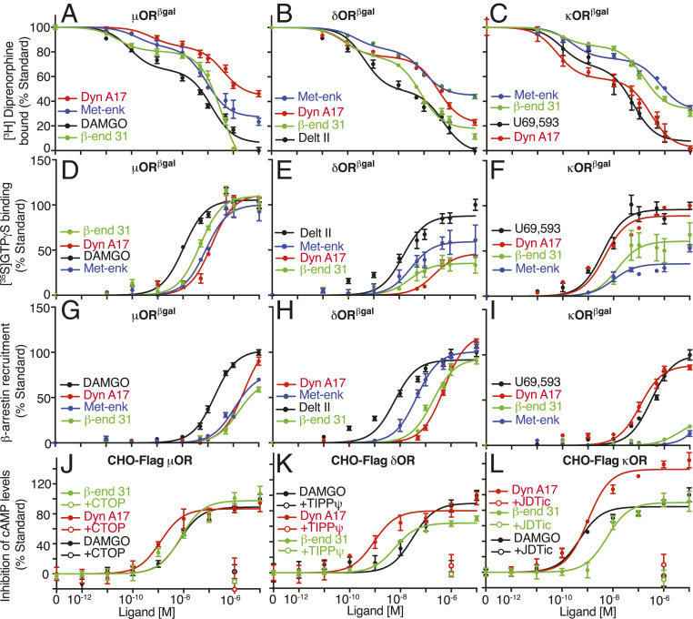
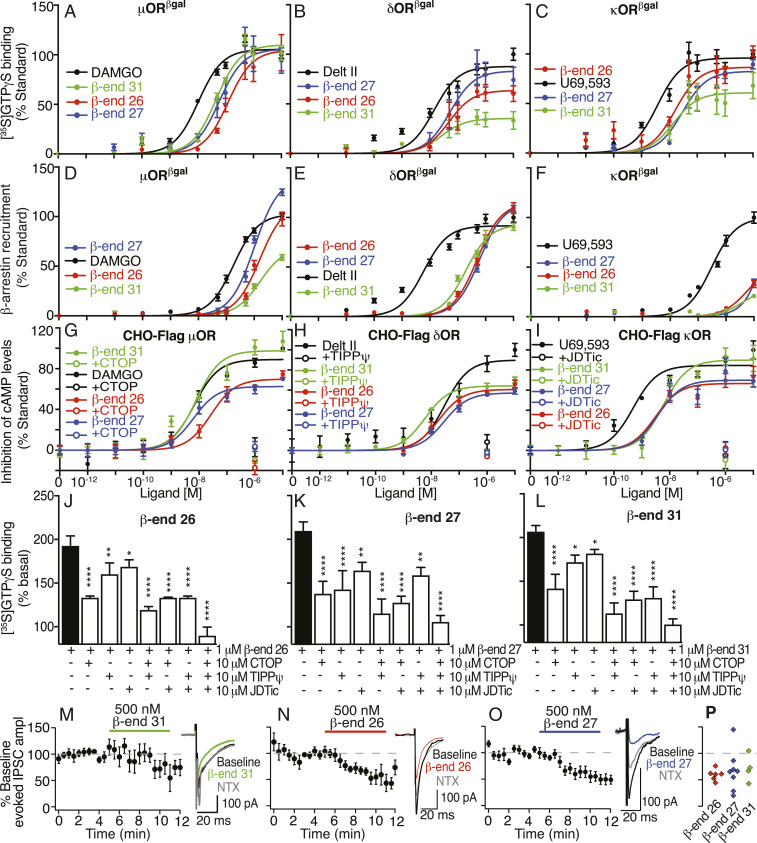
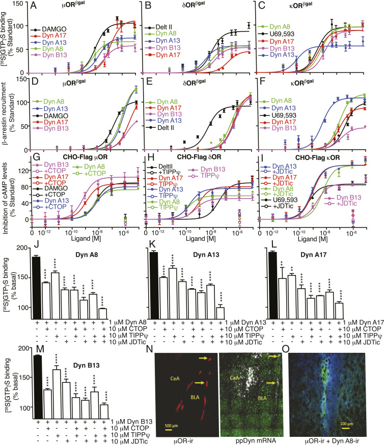
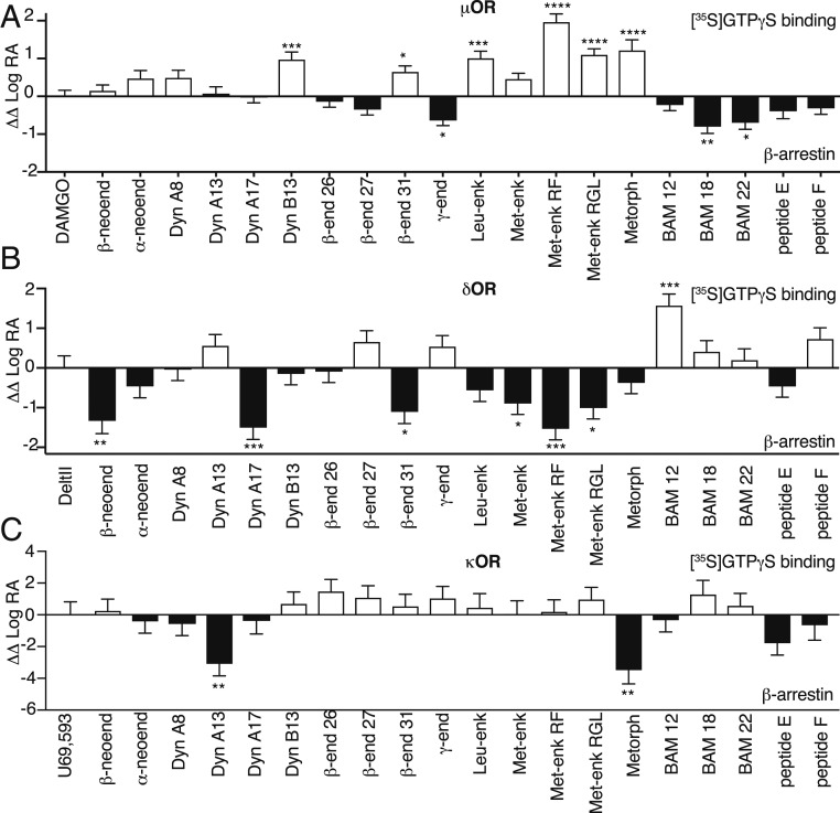
Opioids, such as morphine and fentanyl, are widely used for the treatment of severe pain; however, prolonged treatment with these drugs leads to the development of tolerance and can lead to opioid use disorder. The "Opioid Epidemic" has generated a drive for a deeper understanding of the fundamental signaling mechanisms of opioid receptors. It is generally thought that the three types of opioid receptors (μ, δ, κ) are activated by endogenous peptides derived from three different precursors: Proopiomelanocortin, proenkephalin, and prodynorphin. Posttranslational processing of these precursors generates >20 peptides with opioid receptor activity, leading to a long-standing question of the significance of this repertoire of peptides. Here, we address some aspects of this question using a technical tour de force approach to systematically evaluate ligand binding and signaling properties ([35S]GTPγS binding and β-arrestin recruitment) of 22 peptides at each of the three opioid receptors. We show that nearly all tested peptides are able to activate the three opioid receptors, and many of them exhibit agonist-directed receptor signaling (functional selectivity). Our data also challenge the dogma that shorter forms of β-endorphin do not exhibit receptor activity; we show that they exhibit robust signaling in cultured cells and in an acute brain slice preparation. Collectively, this information lays the groundwork for improved understanding of the endogenous opioid system that will help in developing more effective treatments for pain and addiction.

Keywords: GPCRs; biased agonism; opioid peptides; opioid receptors.


Conflict of interest statement

The authors declare no competing interests.

Figures

Fig. 1.
Fig. 1.
Representative opioid peptides bind and signal at μOR, δOR, and κOR. (AC) Displacement of [3H]diprenorphine binding by β-end 31, Met-enk, and Dyn A17 in membranes (20 μg) from cells expressing either μβgalOR (A), δβgalOR (B), or κβgalOR (C). (DF) [35S]GTPγS binding in membranes (20 μg) from cells expressing either μβgalOR (D), δβgalOR (E), or κβgalOR (F). (GI) β-Arrestin recruitment in cells expressing either μβgalOR (G), δβgalOR (H), or κβgalOR (I). (JL) Inhibition of cAMP levels in CHO cells expressing Flag epitope-tagged μOR (J), δOR (K), or κOR (L). Antagonists (10 μM) to μOR, CTOP (J), to δOR, TIPPψ (K), and to κOR, JDTic (L) block β-end 31-, Met-enk–, and Dyn A17- (1 μM) mediated inhibition in cAMP levels. DAMGO (μOR), Delt II (δOR), and U69,593 (κOR) were used as standards. Data are mean ± SE from three to six independent experiments.
Fig. 2.
Fig. 2.
β-Endorphin peptides signal at μOR, δOR, and κOR. (AC) [35S]GTPγS binding in membranes (20 μg) from cells expressing either μβgalOR (A), δβgalOR (B), or κβgalOR (C). (DF) β-Arrestin recruitment in cells expressing either μβgalOR (D), δβgalOR (E), or κβgalOR (F). (GI) Inhibition of cAMP levels in CHO cells expressing Flag epitope-tagged μOR (G), δOR (H), or κOR (I). Antagonists to μOR, CTOP (G), to δOR, TIPPψ (H), and to κOR, JDTic (I) block β-end peptide-mediated inhibition in cAMP levels. DAMGO (μOR), Delt II (δOR), and U69,593 (κOR) were used as standards in AI. (JL) Increases in [35S]GTPγS binding by β-end peptides in striatal membranes (20 μg) were completely blocked by a combination of antagonists to μOR (CTOP), δOR (TIPPψ), and to κOR (JDTic). (MP) β-End peptides inhibit evoked synaptic GABAAR-mediated IPSC amplitude (ampl) onto VTA neurons in acute brain slices. Time courses for the effects of 500 nM β-end 31 (M, Left), β-end 26 (N, Left), or β-end 27 (O, Left) responses averaged across neurons are shown. Example IPSC traces show inhibition by 500 nM β-end 31 (M, Right), β-end 26 (N, Right), or β-end 27 (O, Right) and partial reversal by 1 μM naltrexone (NTX). (P) Summary showing the magnitude of the change in evoked IPSC amplitude induced by the endorphin peptides in each neuron tested. Data are mean ± SE from three to six independent experiments for AL and mean ± SE for MP (n = 4 to 8 neurons from three rats each).*P < 0.05; **P < 0.01; ****P < 0.0001, one-way ANOVA.
Fig. 3.
Fig. 3.
Dynorphin peptides signal at μOR, δOR, and κOR. (AC) [35S]GTPγS binding in membranes (20 μg) from cells expressing either μβgalOR (A), δβgalOR (B), or κβgalOR (C). (DF) β-Arrestin recruitment in cells expressing either μβgalOR (D), δβgalOR (E), or κβgalOR (F). (GI) Inhibition of cAMP levels by Dyn A8, Dyn A13, Dyn A17, and Dyn B13 in CHO cells expressing Flag epitope-tagged μOR (G), δOR (H), or κOR (I). Antagonists to μOR, CTOP (G), to δOR, TIPPψ (H), and to κOR, JDTic (I) block Dyn A8-, Dyn A13-, Dyn A17-, or Dyn B13-mediated inhibition in cAMP levels. DAMGO (μOR), Delt II (δOR), and U69,593 (κOR) were used as standards. (JM) Increases in [35S]GTPγS binding by Dyn peptides in striatal membranes (20 μg) are completely blocked by a combination of antagonists to μOR (CTOP), δOR (TIPPψ), and to κOR (JDTic). (N, Left) Low-magnification image of a coronal section through rat amygdala showing high levels of expression of μOR immunoreactivity (μOR-ir; red) in ITC clusters of the basolateral nucleus of amygdala (BLA). μOR-ir is barely visible in the CeA. (N, Right) In situ hybridization autoradiographic imaging of an adjacent section with immunohistochemical staining for the neuronal marker NeuN (green), showing silver grains (white) indicating the presence of ppDYN mRNA; ppDYN mRNA is apparent in many cells in the CeA, and in a few of the ITC cell clusters. Yellow arrows (N) indicate μOR-expressing areas in ITC clusters that also express ppDYN mRNA. (O) Higher-magnification image of an ITC cluster in amygdala showing immunocytochemically detected Dyn A8 expression (blue) in proximity to μOR labeling (green). Data (AM) are mean ± SE from three to six independent experiments. Images (N and O) are representative images from sections obtained from four or more rats. **P < 0.01; ****P < 0.0001, one-way ANOVA.
Fig. 4.
Fig. 4.
Bias plots for endogenous opioid peptides at μOR, δOR, and κOR. Bias analysis for signaling by endogenous opioid peptides at μOR (A), δOR (B), or κOR (C) was performed as described in Materials and Methods. Data are mean ± SE from three to six independent experiments. One-way ANOVA; *P > 0.05; **P > 0.01; ***P > 0.001; ****P > 0.0001.

References

    1. Stein C., Opioid receptors. Annu. Rev. Med. 67, 433–451 (2016). - PubMed
    1. Shenoy S. S., Lui F., Biochemistry, Endogenous Opioids (StatPearls, Treasure Island, FL, 2018). - PubMed
    1. Stein C., New concepts in opioid analgesia. Expert Opin. Investig. Drugs 27, 765–775 (2018). - PubMed
    1. Mansour A., Hoversten M. T., Taylor L. P., Watson S. J., Akil H., The cloned mu, delta and kappa receptors and their endogenous ligands: Evidence for two opioid peptide recognition cores. Brain Res. 700, 89–98 (1995). - PubMed
    1. Höllt V., Opioid peptide processing and receptor selectivity. Annu. Rev. Pharmacol. Toxicol. 26, 59–77 (1986). - PubMed

Publication types