. 2021 Jan;42(1):36-44.
doi: 10.1038/s41401-020-0391-9. Epub 2020 May 11.

An optically active isochroman-2H-chromene conjugate potently suppresses neuronal oxidative injuries associated with the PI3K/Akt and MAPK signaling pathways

Affiliations

An optically active isochroman-2H-chromene conjugate potently suppresses neuronal oxidative injuries associated with the PI3K/Akt and MAPK signaling pathways

Ling-Xue Tao et al. Acta Pharmacol Sin. 2021 Jan.

Abstract

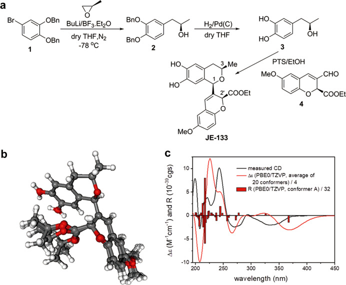
Increasing evidence suggests that the use of potent neuroprotective agents featured with novel pharmacological mechanism would offer a promising strategy to delay or prevent the progression of neurodegeneration. Here, we provide the first demonstration that the chiral nonracemic isochroman-2H-chromene conjugate JE-133, a novel synthetic 1,3-disubstituted isochroman derivative, possesses superior neuroprotective effect against oxidative injuries. Pretreatment with JE-133 (1-10 μM) concentration-dependently prevented H2O2-induced cell death in SH-SY5Y neuroblastoma cells and rat primary cortical neurons. Pretreatment with JE-133 significantly alleviated H2O2-induced apoptotic changes. These protective effects could not be simply attributed to the direct free radical scavenging as JE-133 had moderate activity in reducing DPPH free radical. Further study revealed that pretreatment with JE-133 (10 μM) significantly decreased the phosphorylation of MAPK pathway proteins, especially ERK and P38, in the neuronal cells. In addition, blocking PI3K/Akt pathway using LY294002 partially counteracted the cell viability-enhancing effect of JE-133. We conclude that JE-133 exerts neuroprotection associated with dual regulative mechanisms and consequently activating cell survival and inhibiting apoptotic changes, which may provide important clues for the development of effective neuroprotective drug lead/candidate.

Keywords: MAPK pathway; PI3K/Akt pathway; SH-SY5Y neuroblastoma cells; apoptosis; neurodegeneration; oxidative stress.


Conflict of interest statement

The authors declare no competing interests.

Figures

Fig. 1
Fig. 1
Synthesis and stereochemical analysis of the compound JE-133. a Synthesis of the compound JE-133. b Twenty computed low-energy overlapping solution conformers (level of DFT optimization: CAM-B3LYP/TZVP PCM/MeCN) of (1R,3R,2’S)-JE-133 with an overall Boltzmann population of 87.4%. c Experimental ECD spectrum of JE-133 (black curve) compared with the Boltzmann-weighted PBE0/TZVP (PCM/MeCN) ECD spectrum (red curve) of (1R,3R,2’S)-JE-133 computed for the B97D/TZVP PCM/MeCN conformers. The bars represent the rotational strengths of the lowest-energy conformer
Fig. 2
Fig. 2
Effects of JE-133 on H2O2-induced cytotoxicity in neuronal cells. Cells were pretreated with different concentrations of JE-133 (1, 3 or 10 μM) or NAC (100 μM) for 2 h and then exposed to H2O2 for 24 h. The morphology of SH-SY5Y cells (a) and primary cortical neurons (c) observed under a microscope at 100× magnification after different treatments. Scale bar = 100 μm. The viability of SH-SY5Y cells (b) and primary cortical neurons (d) was determined using the MTT assay, n = 4. The data are presented as the mean ± SEM. ###P < 0.001 vs the control group; *P < 0.05, **P < 0.01, ***P < 0.001 vs the H2O2 group
Fig. 3
Fig. 3
Effects of JE-133 on H2O2-induced apoptosis in SH-SY5Y cells and on free radical scavenging. Cells were pretreated with various concentrations of JE-133 or 100 μM NAC for 2 h and then exposed to H2O2 for 24 h. a Representative FACS analyses of apoptosis. Q1: necrotic cells; Q2: late apoptotic cells; Q3: early apoptotic cells; Q4: live cells. The apoptosis rate is equal to the sum of Q2 and Q3. b A histogram of the percentage of apoptotic cells, n = 4. c Statistical results of cleaved caspase-3. The protein levels of caspase-3 and cleaved caspase-3 were detected by Western blotting using β-actin as a loading control, n = 3. d The direct radical scavenging capacity of FAN was determined using the DPPH free radical scavenging assay, n = 3. The data are presented as the mean ± SEM. ##P < 0.01 vs the control group; *P < 0.05, **P < 0.01 vs the H2O2 group
Fig. 4
Fig. 4
Effects of JE-133 on the phosphorylation of MAPK pathway proteins in H2O2-exposed neuronal cells. Cells were pretreated with JE-133 (10 μM) for 2 h and then exposed to H2O2 for 6 h. The protein levels of p-ERK1/2 (a), p-P38 (b), and p-JNK (c) in SH-SY5Y cells, n = 3. The protein levels of p-ERK1/2 (d), p-P38 (e), and p-JNK (f) in primary cortical neurons, n = 4. The above protein levels were measured by Western blotting using β-actin as a loading control. The data are presented as the mean ± SEM. ##P < 0.01, ###P < 0.001 vs the control group; *P < 0.05 vs the H2O2 group
Fig. 5
Fig. 5
The PI3K/Akt pathway mediated the neuroprotective effect of JE-133 in H2O2-exposed neuronal cells. a The level of p-AKT detected by Western blotting, n = 3. b The effect of LY294002 on the H2O2-induced reduction in viability of SH-SY5Y cells, n = 3. c The effect of LY294002 on the H2O2-induced reduction in viability of primary cortical neurons, n = 4. Cell viability was detected by the MTT assay. The data are presented as the mean ± SEM. #P < 0.05, ###P < 0.001 vs the control group; *P < 0.05, ***P <0.001 vs the H2O2 group; &P < 0.05 vs the H2O2 + JE-133 group
Fig. 6
Fig. 6
Effect of LY294002 on suppressive effect of JE-133 on the phosphorylation of MAPK pathway proteins in H2O2-exposed SH-SY5Y cells. a The protein levels of p-P38 and P38. b The protein levels of p-ERK1/2 and ERK1/2. c The protein levels of p-JNK and JNK. The above protein levels were analyzed by Western blotting using β-actin as a loading control. A histogram of the protein expression ratio of p-P38 (p-ERK1/2, p-JNK)/β-actin based on the results of six independent experiments. The data are presented as the mean ± SEM. #P < 0.05, ##P < 0.01 vs the control group; *P < 0.05, ***P < 0.001 vs the H2O2 group; &P < 0.05, &&P < 0.01 vs the H2O2 + JE-133 group
Fig. 7
Fig. 7
Schematic diagram of the beneficial effects of JE-133 against H2O2-induced neurotoxicity and the potential mechanisms. H2O2 exposure induces intracellular oxidative stress, which triggers the overphosphorylation of the MAPK pathway proteins ERK1/2, P38 and JNK, and induces the upregulation of Akt phosphorylation, consequently activating neuronal apoptosis and ultimately leading to neuronal damage. JE-133 prevents H2O2-induced neuronal damage, which might be mediated by the downregulation of ERK1/2 and P38 phosphorylation and associated with the PI3K/Akt pathway. The dual regulatory effects on the PI3K/Akt and MAPK pathways might synergistically contribute to the neuroprotective effects of JE-133

References

    1. Zhao Y, Zhao B. Oxidative stress and the pathogenesis of Alzheimer’s disease. Oxid Med Cell Longev. 2013;2013:316523. - PMC - PubMed
    1. Poprac P, Jomova K, Simunkova M, Kollar V, Rhodes CJ, Valko M. Targeting free radicals in oxidative stress-related human diseases. Trends Pharmacol Sci. 2017;38:592–607. - PubMed
    1. Jiang T, Sun Q, Chen S. Oxidative stress: a major pathogenesis and potential therapeutic target of antioxidative agents in Parkinson’s disease and Alzheimer’s disease. Prog Neurobiol. 2016;147:1–19. - PubMed
    1. Wang X, Wang W, Li L, Perry G, Lee HG, Zhu X. Oxidative stress and mitochondrial dysfunction in Alzheimer’s disease. Biochim Biophys Acta. 2014;1842:1240–7. - PMC - PubMed
    1. Li J, O W, Li W, Jiang ZG, Ghanbari HA. Oxidative stress and neurodegenerative disorders. Int J Mol Sci. 2013;14:24438–75. - PMC - PubMed

MeSH terms