. 2020 May;4(5):507-517.
doi: 10.1038/s41551-020-0555-4. Epub 2020 May 11.

A co-formulation of supramolecularly stabilized insulin and pramlintide enhances mealtime glucagon suppression in diabetic pigs

Affiliations

A co-formulation of supramolecularly stabilized insulin and pramlintide enhances mealtime glucagon suppression in diabetic pigs

Caitlin L Maikawa et al. Nat Biomed Eng. 2020 May.

Abstract

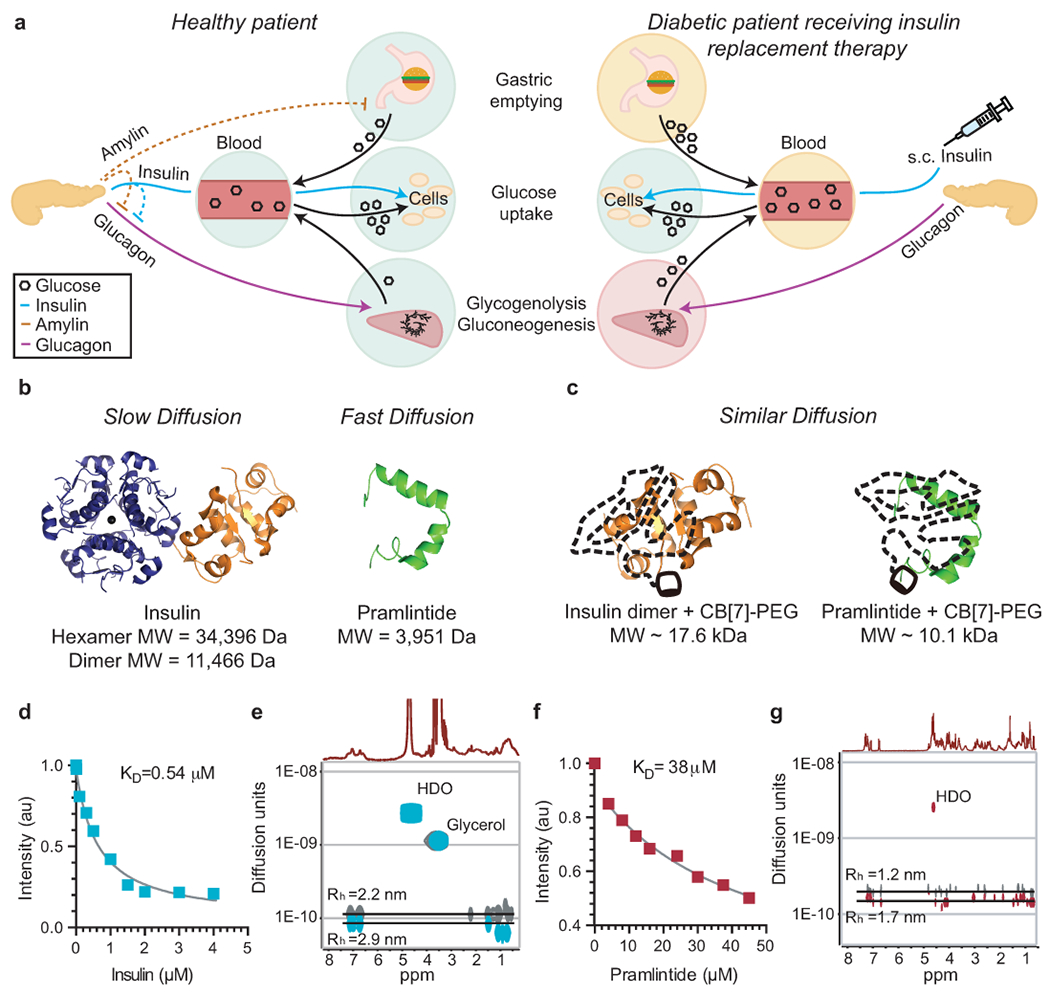
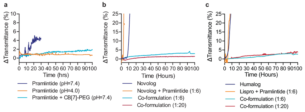
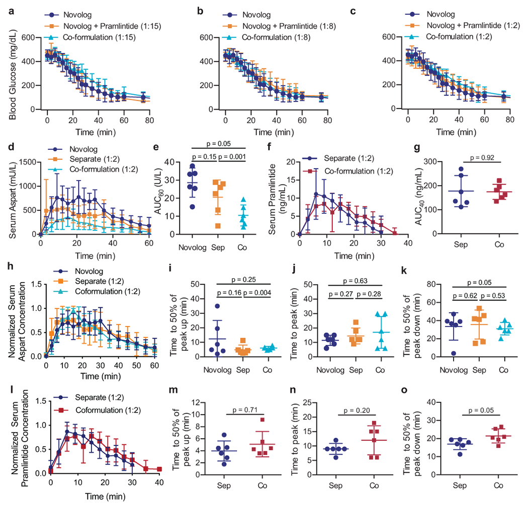
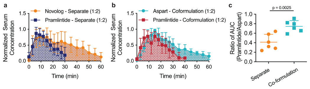
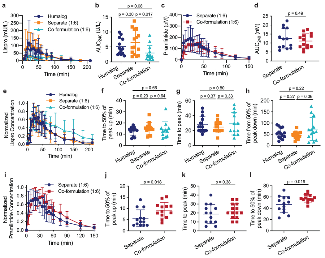
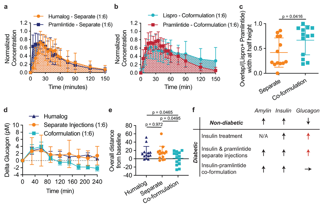
Treatment of patients with diabetes with insulin and pramlintide (an amylin analogue) is more effective than treatment with insulin only. However, because mixtures of insulin and pramlintide are unstable and have to be injected separately, amylin analogues are only used by 1.5% of people with diabetes needing rapid-acting insulin. Here, we show that the supramolecular modification of insulin and pramlintide with cucurbit[7]uril-conjugated polyethylene glycol improves the pharmacokinetics of the dual-hormone therapy and enhances postprandial glucagon suppression in diabetic pigs. The co-formulation is stable for over 100 h at 37 °C under continuous agitation, whereas commercial formulations of insulin analogues aggregate after 10 h under similar conditions. In diabetic rats, the administration of the stabilized co-formulation increased the area-of-overlap ratio of the pharmacokinetic curves of pramlintide and insulin from 0.4 ± 0.2 to 0.7 ± 0.1 (mean ± s.d.) for the separate administration of the hormones. The co-administration of supramolecularly stabilized insulin and pramlintide better mimics the endogenous kinetics of co-secreted insulin and amylin, and holds promise as a dual-hormone replacement therapy.


Conflict of interest statement

Competing interests

E.A.A., B.A.B., D.M.M., C.L.M., and G.A.R. are inventors on a patent filing describing the work reported in this manuscript.

Figures

Fig. 1 |
Fig. 1 |. CB[7]-PEG binds to insulin and pramlintide and alters diffusion rates in formulation.
Scheme of post-mealtime metabolic signaling pathways in a, non-diabetic people and type 1 diabetic people receiving insulin replacement therapy. In non-diabetic people, endogenous insulin promotes cellular glucose uptake and acts with amylin to locally suppress post-prandial glucagon, thus decreasing glycogenolysis & gluconeogenesis. In contrast, treatment of diabetic patients with s.c. insulin alone cannot restore glucagon suppression. Amylin replacement is critical to fully restore metabolic signaling and constitute a true hormone replacement therapy. b, c, Scheme demonstrating how molecular weight affects diffusion rates, which directly impacts absorption kinetics following s.c. administration. b, Standard insulin formulations comprise a mixture of insulin aggregation states (i.e., hexamers and dimers) that exhibit extended duration of insulin action when injected into the s.c. space. In contrast, the pramlintide monomer is rapidly absorbed into the blood. c, After complexation with CB[7]-PEG such that only insulin dimers exist in formulation, insulin and pramlintide have more similar molecular weights and diffusion rates to one another. Acridine orange competitive binding assay of d, aspart (n=1 independent experiment) and f, pramlintide (n=1 independent experiment), indicating binding of CB[7] to both proteins. Diffusion-ordered NMR Spectroscopy (DOSY) provides insight into the formation of protein/CB[7]-PEG complexes and their rates of diffusion in formulation. In these studies, e, Aspart/CB[7]-PEG complex (cyan) exhibits a 30% reduction in diffusion rate when compared to standard dimeric aspart (grey). Moreover, g, Pramlintide/CB[7]-PEG complex (red) exhibits approximately a two-fold reduction in diffusion rate when compared to pramlintide alone (grey). Complexation of the two proteins with CB[7]-PEG results in a ratio in diffusion rates of pramlintide/CB[7]-PEG to aspart/CB[7]-PEG of only 1.6, compared to 2.3 for pramlintide and aspart in typical formulations, indicating the proteins have more similar diffusivities in co-formulation.
Fig. 2 |
Fig. 2 |. Formulation with CB[7]-PEG stabilizes a co-formulation of Novolog or Humalog and pramlintide at physiological pH.
a, In vitro stability of pramlintide formulations at various pH values with and without CB[7]-PEG. b, In vitro stability of pramlintide-aspart (1:6 and 1:20 molar ratio) co-formulations with CB[7]-PEG at physiological pH. c, In vitro stability of pramlintide-lispro (1:6 and 1:20 molar ratio) co-formulations with CB[7]-PEG at physiological pH. Co-formulations were compared to controls of commercial Novolog or Humalog, and mixtures of the incompatible aspart+pramlintide or lispro+pramlintide in the absence of CB[7]-PEG. These assays assess the aggregation of proteins in formulation over time during stressed aging (i.e., continuous agitation at 37ºC) by monitoring changes in transmittance at 540nm. These experiments demonstrate that formulation with CB[7]-PEG prevents protein aggregation over the 100h period assayed, even when commercial formulations aggregate within 10h. Data shown are average transmittance traces for n = 3 samples per group.
Fig. 3 |
Fig. 3 |. Aspart and pramlintide pharmacokinetics following different administration routes in diabetic rats.
Fasted diabetic male rats (n=6) received subcutaneous administration of therapies comprising either (i) commercial Novolog, (ii) commercial Novolog and pramlintide (pH=4) delivered in separate injections, or (iii) aspart-pramlintide co-formulation with CB[7]-PEG. All treatment groups received 1.5U/kg of insulin. Blood glucose levels were evaluated at several ratios of pramlintide to aspart: a, 1:15, b, 1:8, c, 1:2. All pharmacokinetic studies were evaluated with pramlintide at 1:2 aspart:pramlintide ratio. Pharmacokinetics of d, insulin aspart in mU/L or f, pramlintide in ng/mL. The area under the pharmacokinetic curves (AUC) of e, aspart (**p=0.0012) and g, pramlintide for the first 60 minutes or 40 minutes, respectively, after subcutaneous injection. Pharmacokinetics for each rat were individually normalized to peak serum and normalized values were averaged for h, aspart or l, pramlintide concentration for each treatment group. Time to reach 50% of peak i, aspart or m, pramlintide serum concentration (onset). Time to reach peak j, aspart or n, pramlintide serum concentration. Time for k, aspart or o, pramlintide depletion to 50% of peak serum concentration (*p=0.047). Error bars indicate mean ± s.d. with n=6 animals for all groups. Statistical significance was determined by a two-tailed student’s t-test.
Fig. 4 |
Fig. 4 |. Administration of aspart and pramlintide as a co-formulation in diabetic rats enhances pharmacokinetic overlap.
Mean normalized serum concentration (normalized for each individual rat) of Novolog and Pramlintide when administered as a, two separate injections or b, pramlintide-aspart co-formulation with CB[7]-PEG at physiologic pH. c, Ratio of the area under the curve (AUC) of the pharmacokinetic profiles of pramlintide and aspart for administration as separate injections and as a co-formulation (**p=0.0025). Error bars indicate mean ± s.d. with n=6 animals for all groups. Statistical significance was determined by a two-tailed student’s t-test.
Fig. 5 |
Fig. 5 |. Lispro and pramlintide pharmacokinetics following different administration routes in diabetic pigs.
Diabetic female pigs received subcutaneous administration of therapies comprising either (i) commercial Humalog, (ii) commercial Humalog and pramlintide (pH=4) delivered in separate injections, or (iii) lispro-pramlintide co-formulation with CB[7]-PEG. Treatments were administered simultaneously with a 200g meal. All treatment groups received 4U insulin and pramlintide groups received a molar ratio of 1:6 pramlintide to lispro. Pharmacokinetics of a, insulin lispro in mU/L lispro (Humalog n=14; Separate n=15; Co-formulation n=13) or c, pramlintide in pM (Separate n=14; Co-formulation n=14). The area under the pharmacokinetic curves (AUC) of b, lispro (Humalog n=12; Separate n=14; Co-formulation n=13) and d, pramlintide (Separate n=13; Co-formulation n=14) for the first 240 minutes after subcutaneous injection. Pharmacokinetics for each pig were individually normalized to peak concentrations and normalized values were averaged for e, lispro (Humalog n=14; Separate n=15; Co-formulation n=13) or i, pramlintide (Separate n=14; Co-formulation n=14) concentration for each treatment group. Time to reach 50% of peak f, lispro (Humalog n=13;Separate n=14; Co-formulation n=12) or j, pramlintide concentration (onset) (Separate n=13; Co-formulation n=13). Time to reach peak g, lispro (Humalog n=14; Separate n=14; Co-formulation n=12) or k, pramlintide concentration (Separate n=13; Co-formulation n=14). Time for h, lispro (Humalog n=14; Separate n=15; Co-formulation n=13) or l, pramlintide depletion to 50% of peak concentration (Separate n=13; Co-formulation n=13). The specified sample size n refers to a cohort of 11 pigs who received each treatment group an equal number of times. Error bars, mean ± s.d. The Grubbs’ outlier test (alpha=0.05) was used to remove outliers. Statistical significance was determined by a two-tailed student’s t-test.
Fig. 6 |
Fig. 6 |. Overlap between pharmacokinetic curves of lispro and pramlintide and glucagon suppression following treatment with different formulations in diabetic pigs.
Pharmacokinetics of lispro and pramlintide after injection with Humalog and pramlintide as separate injections and as a lispro-pramlintide co-formulation. Mean normalized concentration (normalized individually for each pig) of lispro and pramlintide when administered as a, two separate injections (Humalog n=15; Pramlintide n=13) or b, as a co-formulation (Lispro n=13; Pramlintide n=14) with CB[7]-PEG. The overlap between curves was evaluated as the time during which both lispro and pramlintide concentrations were greater than 0.5 (width at half peak height), shown as a ratio of c, overlap time over the total width of both peaks (Overlap/(Lispro + Pramlintide - Overlap)) (Separate n=13; Co-formulation n=13). Pharmacokinetics of glucagon after a meal and treatment with Humalog alone, Humalog and pramlintide as separate injections or as a lispro-pramlintide co-formulation. Glucagon is plotted as d, change in glucagon concentrations from baseline over 4-hours following treatment administration e, overall distance from baseline by treatment group (sum of individual points) (Humalog n=13; Separate n=14; Co-formulation n=14). The co-formulation reduced glucagon levels compared to Humalog (*p=0.0465) and separate administrations of Humalog and pramlintide (*p=0.0495). f, A summary schematic of how treatment affects post-prandial glucagon. The specified sample size n refers to a cohort of 11 pigs who received each treatment group an equal number of times. Error bars indicate mean ± s.d. The ROUT test (Q=1%) was used to remove outliers. Statistical significance was determined by a two-tailed student’s t-test.

Comment in

Similar articles

Cited by

References

    1. WHO. Diabetes: Key Facts. World Health Organization; (2017).
    1. Borm AK et al. The effect of pramlintide (amylin analogue) treatment on bone metabolism and bone density in patients with type 1 diabetes mellitus. Horm. Metab. Res 31, 472–475 (1999). - PubMed
    1. Gottlieb A et al. Pramlintide as an adjunct to insulin therapy improved glycemic and weight control in people with type 1 diabetes during treatment for 52 weeks. Diabetes. 49, A109–A109 (2000).
    1. Ryan GJ, Jobe LJ & Martin R Pramlintide in the treatment of type 1 and type 2 diabetes mellitus. Clin. Ther 27, 1500–1512 (2005). - PubMed
    1. Edelman S et al. A double-blind, placebo-controlled trial assessing pramlintide treatment in the setting of intensive insulin therapy in type 1 diabetes. Diabetes Care. 29, 2189–2195 (2006). - PubMed

Publication types

MeSH terms