Review
. 2021 Sep;109(3):257-276.
doi: 10.1007/s00223-020-00702-6. Epub 2020 May 11.

The Genetic Epidemiology of Joint Shape and the Development of Osteoarthritis

Affiliations
Review

The Genetic Epidemiology of Joint Shape and the Development of Osteoarthritis

J Mark Wilkinson et al. Calcif Tissue Int. 2021 Sep.

Abstract

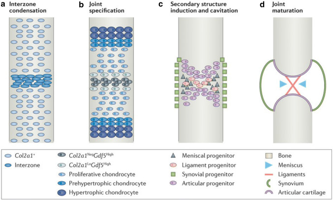
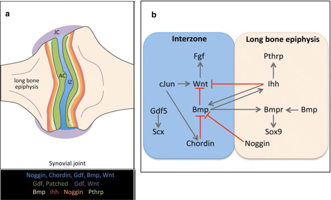
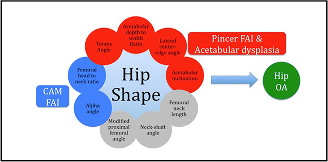
Congruent, low-friction relative movement between the articulating elements of a synovial joint is an essential pre-requisite for sustained, efficient, function. Where disorders of joint formation or maintenance exist, mechanical overloading and osteoarthritis (OA) follow. The heritable component of OA accounts for ~ 50% of susceptible risk. Although almost 100 genetic risk loci for OA have now been identified, and the epidemiological relationship between joint development, joint shape and osteoarthritis is well established, we still have only a limited understanding of the contribution that genetic variation makes to joint shape and how this modulates OA risk. In this article, a brief overview of synovial joint development and its genetic regulation is followed by a review of current knowledge on the genetic epidemiology of established joint shape disorders and common shape variation. A summary of current genetic epidemiology of OA is also given, together with current evidence on the genetic overlap between shape variation and OA. Finally, the established genetic risk loci for both joint shape and osteoarthritis are discussed.

Keywords: Epidemiology; Genetics; Joint development; Joint shape; Osteoarthritis.


Figures

Fig. 1
Fig. 1
Synovial joint development in the mouse. Longitudinal views depicting key steps in the formation of the knee joint. a The first sign of a presumptive joint is a condensation of Col2 + limb bud progenitors at the presumptive joint site; b Joint specification is marked by induction of Gdf5 in the interzone and downregulation of Col2a1; c A joint space is formed by cavitation after progenitors for a variety of secondary joint structures are specified from the Gdf5 + progenitor pool; d Maturation of the synovial joint of the knee occurs during development and early postnatal life. Reproduced from Salazar et al. [165], with permission
Fig. 2
Fig. 2
Spatial expression patterns (a) and principal signaling pathways (b) in synovial joint development. The expression domains of critical signalling pathway components are regionally restricted during development of synovial joints. AC articular cartilage, IZ interzone, JC joint capsule. Reproduced from Salva et al. [12], with permission
Fig. 3
Fig. 3
Disorders of the growing hip joint. a Bilateral hip dysplasia in a skeletally mature individual. The acetabuli are typically shallow and steep, and there is extrusion of the femoral heads with evidence of decreased lateral coverage (arrows); b Perthes’ disease of the right hip in a skeletally mature individual. The right femoral head is broad and flattened and the corresponding acetabululm is similarly shaped (arrow). c Slipped capital femoral epiphysis of the left hip in an adolescent male. The epiphysis has slipped posteromedially from the physis (arrow); d Bilateral cam lesions in a skeletally maturing male, arrows show lateral head-neck junction prominence; e Bilateral pincer lesions in a skeletally mature male, arrows show over-coverage of femoral head
Fig. 4
Fig. 4
Acetabular and proximal femoral morphological parameters associated with OA risk. a HTE horizontal toit externe, SA Sharp’s angle, and AT acetabular tilt; b AIDWR acetabular index of depth to width ratio; c LCEA lateral centre-edge angle and AA alpha angle; d FHFNR femoral head to femoral neck ratio and FNLWR femoral neck length to width ratio; e MPFA modified proximal femoral angle and FNSA femoral neck-shaft angle
Fig. 5
Fig. 5
The biomechanical consequences of non-normal hip shape. Illustration shows relationship between out of range hip shape parameters and the development of osteoarthritis
Fig. 6
Fig. 6
Effect size and risk allele frequency of published osteoarthritis genetic risk loci. Each circle represents a published osteoarthritis risk single-nucleotide variant plotted with its odds ratio (OR; y axis) as a function of the risk allele frequency (x-axis). The different size and colour of each circle shows the sibling relative risk ratio (λs) and the percentage of variance explained on the liability scale respectively (h2L; calculated assuming 13.5% prevalence of OA). Gene annotations are taken from the Ensembl genome browser (human assembly GRCh37). The majority are common variants with small (OR < 2.0) effect sizes, only those with OR > 2.0 are named here

References

    1. Disease GBD, Injury I, Prevalence C. Global, regional, and national incidence, prevalence, and years lived with disability for 310 diseases and injuries, 1990–2015: a systematic analysis for the Global Burden of Disease Study 2015. Lancet. 2016;388:1545–1602. doi: 10.1016/S0140-6736(16)31678-6. - DOI - PMC - PubMed
    1. Global Burden of Disease Study C Global, regional, and national incidence, prevalence, and years lived with disability for 301 acute and chronic diseases and injuries in 188 countries, 1990–2013: a systematic analysis for the Global Burden of Disease Study 2013. Lancet. 2015;386:743–800. doi: 10.1016/S0140-6736(15)60692-4. - DOI - PMC - PubMed
    1. Nuesch E, Dieppe P, Reichenbach S, Williams S, Iff S, Juni P. All cause and disease specific mortality in patients with knee or hip osteoarthritis: population based cohort study. BMJ. 2011;342:d1165. doi: 10.1136/bmj.d1165. - DOI - PMC - PubMed
    1. Yelin E, Murphy L, Cisternas MG, Foreman AJ, Pasta DJ, Helmick CG. Medical care expenditures and earnings losses among persons with arthritis and other rheumatic conditions in 2003, and comparisons with 1997. Arthritis Rheum. 2007;56:1397–1407. doi: 10.1002/art.22565. - DOI - PMC - PubMed
    1. Murphy LB, Cisternas MG, Pasta DJ, Helmick CG, Yelin EH. Medical expenditures and earnings losses among US adults with arthritis in 2013. Arthritis Care Res (Hoboken) 2018;70:869–876. doi: 10.1002/acr.23425. - DOI - PubMed