. 2020 Aug;72(4):513-525.
doi: 10.1007/s10616-020-00397-5. Epub 2020 May 11.

Assessment of proliferation, migration and differentiation potentials of bone marrow mesenchymal stem cells labeling with silica-coated and amine-modified superparamagnetic iron oxide nanoparticles

Affiliations

Assessment of proliferation, migration and differentiation potentials of bone marrow mesenchymal stem cells labeling with silica-coated and amine-modified superparamagnetic iron oxide nanoparticles

Dong Yao et al. Cytotechnology. 2020 Aug.

Abstract

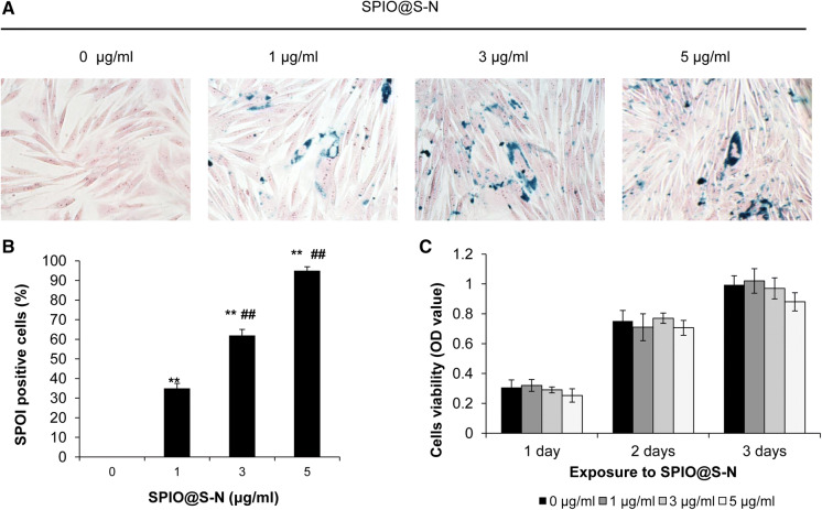
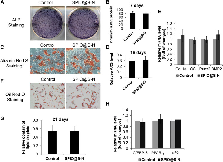
Superparamagnetic iron oxide nanoparticles have been widely used for cell labeling in preclinical and clinical studies, to improve labeling efficiency, particle conjugation and surface modifications are developed, but some modified SPIONs exert side-effect on physiological activity of cells, which cannot be served as ideal cell tracker. In this study, amine-modified silica-coated SPIO (SPIO@SiO2-NH2, SPIO@S-N) nanoparticles were used to label bone marrow derived mesenchymal stem cells (BM-MSCs), then the stem cell potentials were evaluated. It was found BM-MSCs could be efficiently labeled by SPIO@S-N nanoparticles. After labeling, the BM-MSCs viability kept well and the migration ability increased, but the osteogenesis and adipogenesis potentials were not impaired. In steroid associated osteonecrosis (SAON) bone defect model, stem cell implantation was performed by injection of SPIO@S-N labeled BM-MSCs into marrow cavity locally, it was found the SPIO positive cells homed to the periphery of defect region in control group, but were recruited to the defect region in poly lactic-coglycolic acid/tricalcium phosphate (PLGA/TCP) scaffold implantation group. In conclusion, SPIO@S-N nanoparticles promoted migration while retained proliferation and differentiation ability of BM-MSCs, implying this kind of nanoparticles could be served not only an ideal tracking marker but also an accelerator for stem cell homing during tissue repair.

Keywords: Differentiation; Mesenchymal stem cells; Migration; Steroid associated osteonecrosis; Superparamagnetic iron oxide nanoparticles; Viability.


Conflict of interest statement

The authors have no financial conflict of interest.

Figures

Fig. 1
Fig. 1
Rabbit BM-MSCs labeled with SPIO@SiO2-NH2 nanoparticles and the cell viability. a Rabbit BM-MSCs were labeled by SPIO@S-N nanoparticles with the concentration of 0 μg/ml, 1 μg/ml, 3 μg/ml and 5 μg/ml respectively for 16 h, and then Prussian blue staining was performed (SPIO@S-N: SPIO@SiO2-NH2). b Quantification of SPIO@S-N nanoparticles positive labeled cells in a. (3 independent experiments, 10 regions (25 cells in each region) were chosen for cell counting, **p < 0.01 vs. control group; ##p < 0.01 vs. other group). c Rabbit BM-MSCs were incubated with SPIO@S-N nanoparticles (0, 1, 3 and 5 μg/ml) for 1, 2 and 3 days respectively, and then CKK-8 assay was performed (n = 3)
Fig. 2
Fig. 2
SPIO@SiO2-NH2 nanoparticles labeled Rabbit BM-MSCs migration ability. a Rabbit BM-MSCs with or without SPIO@S-N nanoparticles (5 μg/ml) labeling were cultured in 6-well tissue culture plate for 24 h, then scratched wounds were generated and the cells were incubated in medium containing 5% fetal bovine serum (FBS) for 12 h, microscopic images were captured at time points of hour 0, 6, and 12. b The rate and extent of BM-MSCs migration in a was quantitated by Image pro Plus at time point of 6 h and 12 h (3 independent experiments and 6 randomly selected fields were captured in each sample, *p < 0.05 vs. control group). c Rabbit BM-MSCs incubated with or without SPIO@S-N nanoparticles (5 μg/ml) labeling for 24 h, then mRNA were extracted and qRT-PCR was performed to detect SDF1and CXCR4 gene expressions (n = 5). d BM-MSCs with or without labeling for 24 h were extracted and western blot was performed for SDF-1 and CXCR4 protein expression. e Quantification of immunoblot density of SDF-1 and CXCR4 in d. Statistical significance was defined as **p < 0.01
Fig. 3
Fig. 3
Osteogenic and adipogenic differentiation potentials of SPIO@SiO2-NH2 nanoparticles labeled rabbit BM-MSCs. Primary rabbit MSCs labeled with or without SPIO@S-N nanoparticles (5 µg/ml) were placed into 6-well plate and were incubated for 16 h, then were refreshed with osteogenic induction medium for further culture. a ALP staining was performed at day 7 after induction. b Alkaline phosphatase (ALP) activity was detected at day 7 after induction (n = 5). c Alizarin Red S (ARS) staining was performed at day 14 after induction. d Quantification of relative ARS level in c (n = 5). e RNA extracted at day 7 after induction and qRT-PCR was performed to detect osteogenic differentiation related gene expressions (n = 5). f After adipogenic induction for 21 days, Oil Red O staining was performed to lipid formation ability. g Quantification of relative lipid drops level in f (n = 5). h RNA extracted at day 21 after induction and qRT-PCR was performed to detect adipogenic differentiation related gene expressions (n = 5)
Fig. 4
Fig. 4
SPIO@SiO2-NH2 nanoparticles labeled BM-MSCs tracking in rabbit SAON bone defect model. A Schematic diagram for SAON bone defect model establishment and SPIO@S-N nanoparticles pre-labeled BMSCs PLGA/TCP scaffold implantation. B MRI was performed for proximal femur with a 3.0 T clinical whole-body MR unit by using a knee coil at day 0 and day 7 after implantation, GRET2W MRI images were used for analysis of the target region (yellow arrow indicated MSCs injection site, red arrow indicated bone defect region). C Prussian blue plus nuclear fast red staining of the histological sections of empty control and P/T. I, The location of the bone tunnel was marked with a dotted line (upper, ×5) and the enlarged images in the defect zone (I and III) and peripheral zone of defect (II and IV) respectively. SPIO positive cells were indicated by black arrows (lower, ×20). D SPIO positive cells on the histological sections were counted (n = 10) and plotted. **p < 0.01 compared between P/T group and empty control group, ##p < 0.01 compared between defect zone and peripheral zone in each group

References

    1. Andreas K, Georgieva R, Ladwig M, Mueller S, Notter M, Sittinger M, Ringe J. Highly efficient magnetic stem cell labeling with citrate-coated superparamagnetic iron oxide nanoparticles for MRI tracking. Biomaterials. 2012;33:4515–4525. - PubMed
    1. Askari AT, et al. Effect of stromal-cell-derived factor 1 on stem-cell homing and tissue regeneration in ischaemic cardiomyopathy. Lancet. 2003;362:697–703. - PubMed
    1. Bulte JW, et al. Magnetodendrimers allow endosomal magnetic labeling and in vivo tracking of stem cells. Nat Biotechnol. 2001;19:1141–1147. - PubMed
    1. Chang YK, Liu YP, Ho JH, Hsu SC, Lee OK. Amine-surface-modified superparamagnetic iron oxide nanoparticles interfere with differentiation of human mesenchymal stem cells. J Orthop Res. 2012;30:1499–1506. - PubMed
    1. Chen YC, et al. The inhibitory effect of superparamagnetic iron oxide nanoparticle (Ferucarbotran) on osteogenic differentiation and its signaling mechanism in human mesenchymal stem cells. Toxicol Appl Pharmacol. 2010;245:272–279. - PubMed

LinkOut - more resources