

Deep learning-based fully automatic segmentation of wrist cartilage in MR images
- PMID: 32394453
- PMCID: PMC7784718
- DOI: 10.1002/nbm.4320
Deep learning-based fully automatic segmentation of wrist cartilage in MR images
Abstract
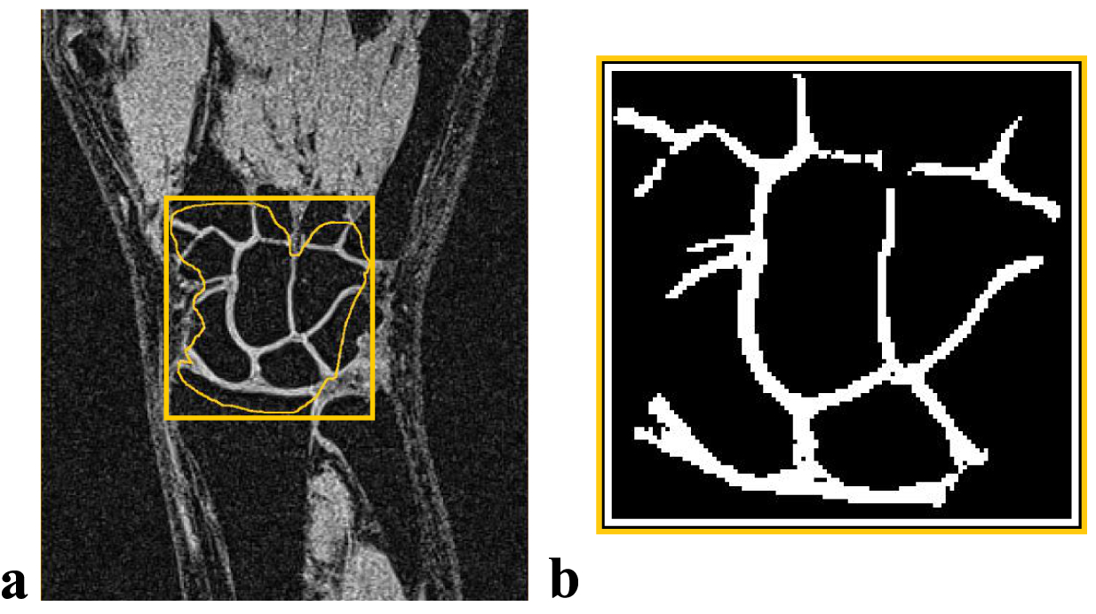
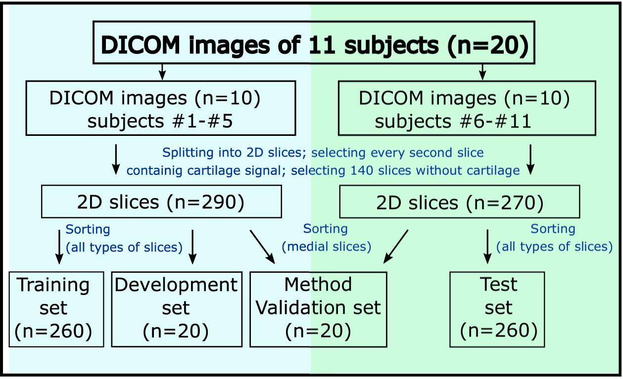
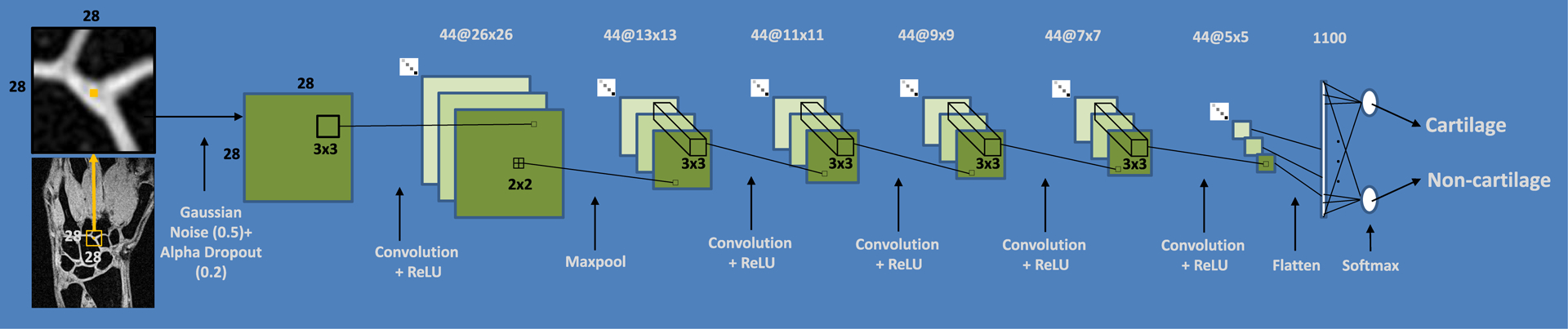
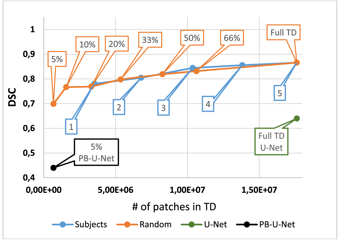
The study objective was to investigate the performance of a dedicated convolutional neural network (CNN) optimized for wrist cartilage segmentation from 2D MR images. CNN utilized a planar architecture and patch-based (PB) training approach that ensured optimal performance in the presence of a limited amount of training data. The CNN was trained and validated in 20 multi-slice MRI datasets acquired with two different coils in 11 subjects (healthy volunteers and patients). The validation included a comparison with the alternative state-of-the-art CNN methods for the segmentation of joints from MR images and the ground-truth manual segmentation. When trained on the limited training data, the CNN outperformed significantly image-based and PB-U-Net networks. Our PB-CNN also demonstrated a good agreement with manual segmentation (Sørensen-Dice similarity coefficient [DSC] = 0.81) in the representative (central coronal) slices with a large amount of cartilage tissue. Reduced performance of the network for slices with a very limited amount of cartilage tissue suggests the need for fully 3D convolutional networks to provide uniform performance across the joint. The study also assessed inter- and intra-observer variability of the manual wrist cartilage segmentation (DSC = 0.78-0.88 and 0.9, respectively). The proposed deep learning-based segmentation of the wrist cartilage from MRI could facilitate research of novel imaging markers of wrist osteoarthritis to characterize its progression and response to therapy.
Keywords: applications, cartilage, human study, methods and engineering, musculoskeletal, postacquisition processing, quantitation.
© 2020 John Wiley & Sons, Ltd.
Conflict of interest statement
The authors declares that there is no conflict of interest
Figures
References
-
- Visser AW, Bøyesen P, Haugen IK, Schoones JW, Van der Heijde DM, Rosendaal FR, Kloppenburg M. Radiographic scoring methods in hand osteoarthritis – a systematic literature search and descriptive review. Osteoarthritis Cartilage 2014;22(10):1710–1723. - PubMed
-
- Link T, Stahl R, Woertler K. Cartilage imaging: Motivation, techniques, current and future significance. Eur Radiol 2007;17(5):1135–1146. - PubMed
-
- Link T. Cartilage Imaging: Significance, Techniques, and New Developments. New York, NY: Springer Science & Business Media; 2011. 93–102 pp.
-
- Eckstein F, Kunz M, Schutzer M, Hudelmaier M, Jackson RD, Yu J, Eaton CB, Schneider MSE. Magnetic resonance imaging (MRI) of articular cartilage in knee osteoarthritis (OA): morphological assessment. Osteoarthritis Cartilage 2006;14:46–75. - PubMed
Publication types
MeSH terms
Grants and funding
LinkOut - more resources
Full Text Sources
Medical
